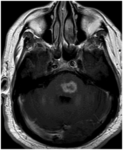
Axial FLAIR through the level of the pons.

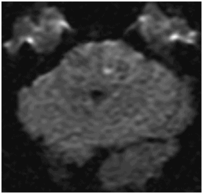
Axial T2WI at the same level.
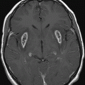
Axial T1WI postcontrast through the level of the pons.
Central Nervous System Blastomycosis
Primary Diagnosis
Central nervous system blastomycosis
Differential Diagnoses
Metastasis
Lymphoma
Sarcoid
Listeria rhombencephalitis
Other causes of rhombencephalitis
Brainstem glioma
Imaging Findings
Fig. 42.1: Axial FLAIR image through the level of the pons demonstrated diffuse swelling of the brainstem, with FLAIR hyperintensity involving the entire pons and bilateral middle cerebellar peduncles, and mass effect to the fourth ventricular floor. Fig. 42.2: Axial T2 image through the same level demonstrated the same findings; however, prominent hypointensity at the center of the lesion was noted. Fig. 42.3: Axial postcontrast T1WI at the same level demonstrated heterogeneous ring enhancement of the T2 hypointense area. Fig. 42.4: DWI through the same level demonstrated a tiny focus of diffusion restriction (with low ADC value, not shown) at the center of the T2 hypointense and relatively less enhancing area of the lesion. Spectroscopy demonstrated high choline-to-creatinine ratio and decreased NAA peak (not shown).
Discussion
In a patient from an endemic area, a gradually worsening disease process involving the brainstem, with diffuse enlargement and FLAIR hyperintensity on imaging, is suggestive for CNS blastomycosis. This is particularly true with the confirmed presence of a central T2 hypointensity that demonstrates heterogeneous ring enhancement in the absence of congruent diffusion restriction. Central T2 hypointensity can often be seen in mucinous metastasis (see Part VI: Case 102), lymphoma, and neurosarcoidosis. In an immunocompetent patient with CNS lymphoma, solid enhancement, and incongruent diffusion, restriction is typically demonstrated on imaging studies, in the absence of heterogeneous ring enhancement. In lymphoma patients, the entire T2 hypointense area usually demonstrates diffusion restriction because of hypercellularity. Ring enhancement is a feature of CNS lymphoma in an immunocompromised patient, or in a patient with prior treatment – not a treatment-naïve CNS lymphoma in an immunocompetent patient. The combination of T2 hypointensity, heterogeneous ring-like enhancement, and extensive perilesional FLAIR hyperintensity can be seen in a metastasis from a mucinous adenocarcinoma, either from the gastrointestinal tract or from the lungs. Although metastasis from these primary sites can involve any regions of the brain, involvement of isolated brainstem is uncommon. Absence of a known primary excludes diagnosis of metastatic disease.
Intraparenchymal CNS sarcoidosis can present with similar imaging findings. However, intraparenchymal lesions are usually associated with adjacent leptomeningeal disease, notably absent in this patient. Additionally, isolated involvement of the brainstem in CNS sarcoidosis is rare. Listeria is the most common cause of rhombencephalitis. The absence of CNS pleocytosis and negative CSF culture findings exclude a diagnosis of CNS listeriosis. Based on the imaging appearance, brainstem gliomas should be considered in the differential diagnosis, particularly in conjunction with the presence of high choline and low NAA levels, as well as the clinical presentation. However, prominent T2 hypointensity and tiny central, rather than peripheral, diffusion restriction is atypical for high-grade brainstem gliomas.
Blastomyces dermatitidis is the causative agent for blastomycosis. Blastomycosis is an uncommon, but potentially serious fungal infection, endemic in the Mississippi and Ohio River basins, and in the Great Lakes and the St Lawrence River regions. Occasionally, it can be found in non-endemic areas as well. There is no predilection for age, sex, race, or occupation, but it occurs more frequently in immunocompromised hosts, particularly in patients with AIDS. It primarily affects the pulmonary system, frequently manifesting as acute or chronic pneumonia. Central nervous system dissemination accounts for 5–10% of extrapulmonary blastomycosis and usually manifests as an intracranial mass lesion, spinal cord abscess, or epidural abscess. Brain masses may be associated with adjacent leptomeningitis; however, presentation with isolated meningitis is rare. The cerebellum is frequently involved, but blastomycosis can involve any region of the brain. Multiple lesions throughout the brain mimicking multiple cerebral metastases have also been described in the literature. Cerebrospinal fluid analysis is not as sensitive as CSF culture in the diagnosis of blastomycosis. Central nervous system blastomycosis is usually treated with amphotericin B, in combination with oral azoles.


Stay updated, free articles. Join our Telegram channel

Full access? Get Clinical Tree


