CASE 42 A 34-year-old woman complains of nonspecific abdominal pain. Fig. 42.1 Axial contrast-enhanced CT image shows an enhancing exophytic splenic lesion (arrow) arising from the inferior pole of the spleen. An axial contrast-enhanced computed tomography (CT) image (Fig. 42.1) demonstrates a well-defined exophytic lesion arising from the inferior pole of the splenic parenchyma and shows homogeneous enhancement after contrast administration. Hemangioma of the spleen Hemangiomas represent the most common primary benign neoplasms of the spleen, being recognized in 0.3 to 14.0% of cases at autopsy. These lesions, which tend to be slightly more common in men, result from the proliferation of splenic vascular channels with endothelial lining and may undergo several changes, including fibrosis, hemorrhage, and necrosis; they usually tend to grow slowly, so that symptoms and complications, although unusual, manifest in late adulthood. The term hemangiomatosis refers to the presence of multiple hemangiomas within the splenic parenchyma; this condition may be part of a systemic angiomatosis as described in Klippel-Trénaunay-Weber, Turner, Kasabach-Merritt, and Beckwith-Wiedemann syndromes.
Clinical Presentation
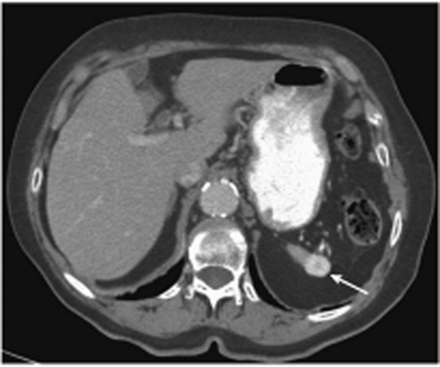
Radiologic Findings
Diagnosis
Differential Diagnosis
Discussion
Background
Clinical Findings
Related posts:


Stay updated, free articles. Join our Telegram channel

Full access? Get Clinical Tree


