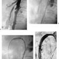
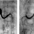
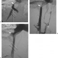
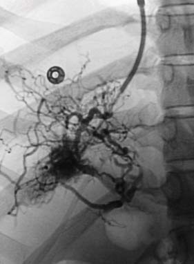
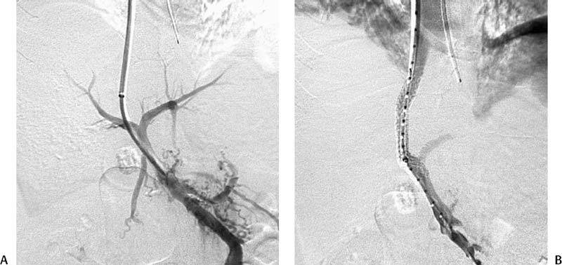
CASE 42 A 51-year-old male was admitted to the gastroenterology service with nausea, vomiting, and abdominal distention. Physical exam revealed ascites and hepatomegaly. Liver enzymes were elevated. Figure 42-1 Hepatic venography from an internal jugular vein approach shows occlusion of the main right and middle hepatic veins and opacification of multiple small hepatic venous collaterals in a spider web pattern. Sonographic evaluation of the right upper quadrant showed occlusion of the right and middle hepatic veins. Conventional venography showed a spider web pattern of innumerable, unnamed hepatic venous branches within the right lobe of the liver (Fig. 42-1). The main right and middle hepatic veins were occluded. Subacute Budd-Chiari syndrome complicated by portal hypertension. Using a Rösch-Uchida transjugular liver access set (Cook, Bloomington, Indiana), a small collateral hepatic vein branch was accessed, the portal vein was punctured, and wire access into the superior mesenteric vein (SMV) was achieved. Venography showed patency of the SMV and portal veins. Using an 8-mm BlueMax balloon catheter (Boston Scientific, Natick, Massachusetts), a tract was dilated through the liver parenchyma. A 10-mm diameter, 6-cm long Viatorr stent (W. L. Gore & Associates, Flagstaff, Arizona) was deployed, extending from the right portal vein to the inferior vena cava (IVC) (Fig. 42-2). Micropuncture system (Cook, Bloomington, Indiana) Amplatz wire (Boston Scientific, Natick, Massachusetts) 9F vascular sheath (Cook, Bloomington, Indiana) Rösch-Uchida Transjugular Liver Access Set (Cook, Bloomington, Indiana) BlueMax 8-mm balloon catheter (Boston Scientific, Natick, Massachusetts) 6-cm Viatorr stent-graft (W. L. Gore & Associates, Flagstaff, Arizona)
Clinical Presentation
Radiologic Studies
Diagnosis
Treatment
Equipment
Discussion
Background
Stay updated, free articles. Join our Telegram channel

Full access? Get Clinical Tree


