Axial T1WI at the level of C1–C2.
Central Nervous System Aspergillosis
Primary Diagnosis
Central nervous system aspergillosis
Differential Diagnoses
Brain metastasis
Pyogenic brain abscesses
Imaging Findings
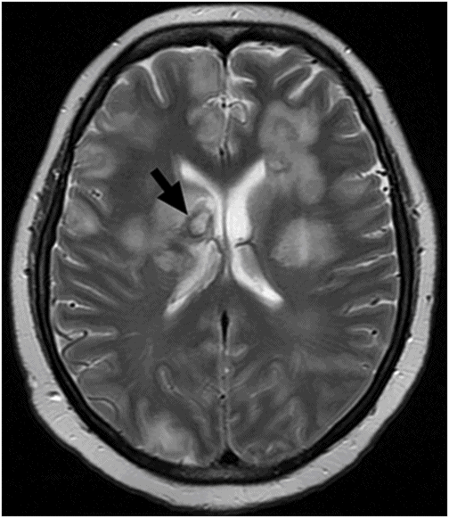
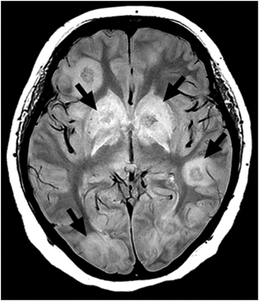
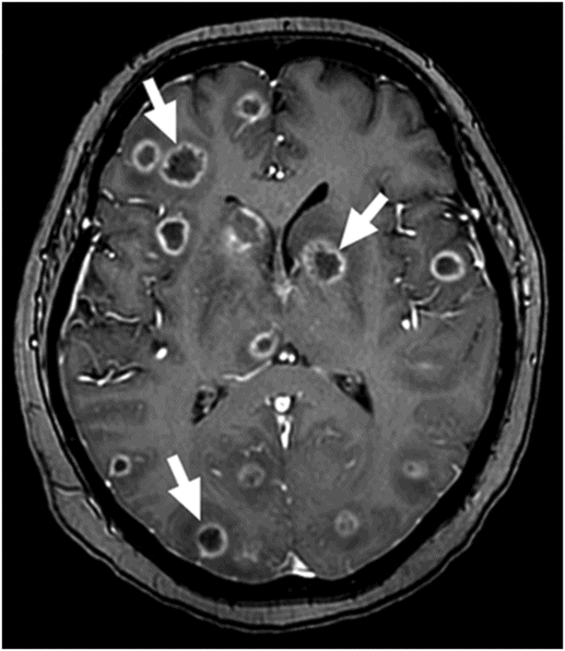
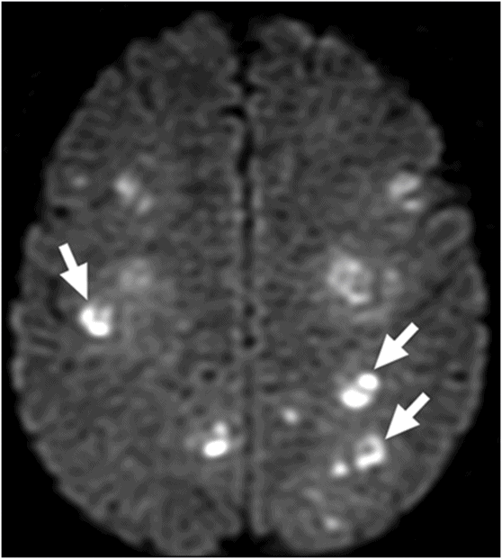
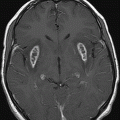
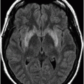
Fig. 43.1: Axial T1WI at the level of C2 demonstrated excessive, subcutaneous fat accumulation in the upper neck, secondary to chronic steroid use. Fig. 43.2: Axial T2WI at the level of the lateral ventricles showed multiple rounded lesions associated with a moderate degree of vasogenic edema. Note that some lesions have a marked hypointense rim, which can represent hemorrhage, or deposition of other paramagnetic substances such as iron, manganese, or magnesium (arrow). Fig. 43.3: Axial proton density image at the level of the basal ganglia showed multiple hyperintense nodules with a moderate degree of vasogenic edema in the basal ganglia and subcortical white matter (arrows). Fig. 43.4: Axial enhanced fat-saturated T1WI at the level of the basal ganglia showed multiple, widespread ring-enhancing lesions in the brain parenchyma (arrows). Fig. 43.5: Axial DWI at the level of the central semiovale demonstrated that the multiple ring-enhancing lesions showed internal restricted diffusion of the molecules’ motion. Apparent diffusion coefficient map (not shown) demonstrated that the areas of increased signal have low values.
Discussion
Imaging findings demonstrating multiple ring-enhancing lesions, with restricted diffusion, hemorrhage, and surrounding vasogenic edema, in a chronically immunosuppressed patient, with rapid neurologic deterioration, are consistent with the presence of an opportunistic infection, such as aspergillosis.
Central nervous system metastases can occur in around 15–40% of patients with malignant cancer. The most common primary metastatic CNS cancers include lung, breast, skin, colon, pancreas, testes, ovary, cervix, renal cell carcinoma, and melanoma. The majority of patients with CNS metastases are asymptomatic (60–75%). When symptomatic, patients may present with headache, seizure, syncope, focal neurologic deficit, or papilledema. Metastasis tends to be located at the gray-white junction and at watershed zones between major arterial vascular territories. Up to 80% of brain metastases occur in the cerebral hemispheres, 15% in the cerebellum, and 3% in the basal ganglia. More rarely, metastatic infiltration of the choroid plexus can include the meninges, ventricular system, pituitary gland, and along blood vessels.
Hemorrhagic metastasis is more commonly associated with melanoma, choriocarcinoma, renal cell carcinoma, and thyroid cancer. Lung and breast metastases are also known to bleed; moreover, they are the most common hemorrhagic metastatic CNS lesions, because of their high prevalence. On MRI, the majority of these lesions are iso- or hypointense on T1, hyperintense on T2, and exhibit an avid solid, or ring of, enhancement. Diffusion-weighted images of hemorrhagic CNS metastases are usually negative, demonstrating restricted diffusion, with high values on ADC maps. Vasogenic edema, usually a common feature, tends to be disproportionate in relation to the size of the lesion. The combination of the patient’s medical history (age, absence of malignancy) and imaging findings of diffuse and multiple restricted diffusion foci makes CNS metastasis an unlikely diagnosis in this case.
Pyogenic brain abscesses are known to have all of the previously mentioned imaging characteristics including ring enhancement, vasogenic edema, signal changes, and typical, internal restricted diffusion. Pyogenic brain abscesses are unquestionably an excellent probable diagnostic possibility in this patient. The clinical history of immunosuppression should raise suspicion for the possibility of an opportunistic infection.
Aspergillus is a ubiquitous saprophytic fungus commonly found in soil, on plants and building materials, as well as in household dust and in decaying organic matter. There are many different species of Aspergillus, but the most common species that can cause aspergillosis are Aspergillus fumigatus and Aspergillus flavus. Affected individuals are usually infected by inhaling aspergillus spores.
The most clinically important forms of aspergillosis are invasive aspergillosis, allergic bronchopulmonary aspergillosis, and aspergillomas. Invasive aspergillosis usually affects immunosuppressed individuals, in which the fungi invade and damage multiple tissues. It more commonly affects the lungs and paranasal sinuses, but can spread throughout the body and cause infection in other organs, such as the brain, its membranes, and other CNS components. Intracranial spread of aspergillosis is a severe and commonly fatal complication, occurring in 10–20% of cases with a very high mortality rate of more than 90%.
Aspergillus can reach the CNS by three different routes. The first is by hematogenous spread from a remote extracranial focus, commonly from the lungs, leading to occlusion of large and middle-sized arteries. The second is by contiguous extension from an extracranial location, commonly from the paranasal sinuses (rhino-cerebral). The third route of CNS entry is iatrogenically, by direct introduction of aspergillus via a surgical intervention. Other potential sites of primary infection include the ear canal, aorta, heart, vertebral disks, and/or cranial bones following traumatic injury or from surgical contamination.
Central nervous system aspergillosis is a worrisome potential complication in patients undergoing chemotherapy or on immunosuppressive therapy following solid organ or bone marrow transplantation. Additional patients at risk for opportunistic CNS aspergillosis include individuals with diabetes, autoimmune diseases requiring steroids, hepatic insufficiency, chronic obstructive pulmonary disease, previous brain lesions, or HIV-AIDS. Previously injured brain tissue (glial scars) may aid the attachment and subsequent invasion of the fungal spores, possibly because of an abnormal blood-brain barrier.
Various imaging patterns can be seen in patients with CNS aspergillosis, such as single or multiple infarcts, single or multiple ring-enhancing lesions (consistent with abscess formation), and dural or vascular infiltration arising from the paranasal sinuses or orbits. Other findings include meningitis, encephalitis, mycotic aneurysm, subdural/epidural abscesses, hemorrhagic transformation of infarcted areas, and sellar abscesses. Signs of hemorrhage may also be seen secondary to vascular invasion.
In order to depict all relevant MRI features in CNS aspergillosis, the protocol should include T1- and T2-weighted sequences, T2*-weighted sequences for an early detection of hemorrhage, DWI to detect early signs of ischemia, and contrast-enhanced T1-weighted images for abscess identification. Spectroscopy may be helpful in analysis of amino acid and lipid levels, and lactate peaks. The presence of multiple peaks seen between 3.6 and 3.8 ppm, due to presence of the disaccharide trehalose, is a distinguishable feature of fungal abscesses.
Diffusion-weighted imaging plays an important role in detecting early manifestations of ischemic CNS lesions because of the angioinvasive character of the fungi, particularly in the hematogenous spread of spores. Involvement of the basal ganglia is a characteristic finding that may indicate a predominant involvement of the lenticulostriate and thalamoperforating arteries. In the majority of the patients, an infarction is followed by a conversion to an infected area, with subsequent abscess formation.
When there is extension of aspergillosis from the paranasal sinuses, initial involvement is seen, in most cases, as a non-specific contrast-enhancing pansinusal mucosal thickening of the sinuses on CT or MRI. Serious complications may exist in cases of infiltration and erosion of the adjacent bony structures, leading to focal meningitis. In cases of invasive lesions in the brain, a well-defined granuloma can usually be seen in the basifrontal or anterior temporal region, often having continuity with the adjacent sinus disease. These granulomas are virtually indistinguishable from tuberculous granulomas or other chronic granulomas.
Aspergillus granulomas are typically T1 hypointense, with a T2 hyperintense center and hypointense wall, attributable to paramagnetic substances such as iron, manganese, and magnesium that are found in the fungal hyphae. Granulomas also show mild to moderate ring-like contrast enhancement and 25% of lesions can be purely hemorrhagic. Dural enhancement is generally seen in lesions adjacent to involved sinuses and the existence of a concomitant sinonasal disease points toward fungal etiology. Aspergillus spores have a predilection for the anterior and middle cranial fossa and abscesses in the cerebellum are extremely rare.
Central nervous system aspergillosis may occur in any age group, with a mean age of 43 years, with a slight male predominance. Regarding signs and symptoms, patients commonly present with headache, fever, mental status changes, generalized seizures, focal neurologic deficits, hemiplegia, vision changes, and cranial nerve palsy. Aspergillus fumigatus is the predominant infectious species found in patients with aspergillosis. It has a propensity to invade the blood vessels following the release of the enzyme elastase, leading to coagulative necrosis, infarcts, mycotic aneurysms, and hemorrhagic lesions. Galactomannan serologic levels are also helpful diagnostic values, rapid diagnostic PCR techniques performed on the CSF and serum could facilitate a faster and simpler diagnostic approach.
Voriconazole has become the standard of care in most cases of CNS aspergillosis. Mortality is high even among patients who received medical treatment, but patients who underwent neurosurgery had a better survival than those who received only medical therapy. Surgical procedures may involve abscess removal or drainage, excision of infected mass, orbital exenterations, debridement of inner ear, and debridement of the skull base. The aim of treatment should be the complete removal of the masses as early as possible.
In patients with invasive aspergillosis, providing intradural extension has not yet occurred, total excision may be curative without systemic use of antifungal agents. However, in patients in whom total removal cannot be achieved or intradural invasion has already been confirmed, intensive systemic therapy with antifungal agents must be administered postoperatively to prevent subsequent lethal vasculitis or meningoencephalitis development.
Incidence of invasive aspergillosis appears to be on the rise. A comprehensive understanding of the infectious process, a high index of suspicion, and advanced laboratory and radiologic diagnostic techniques, may allow early diagnosis.
Stay updated, free articles. Join our Telegram channel

Full access? Get Clinical Tree


