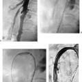
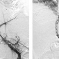
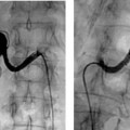
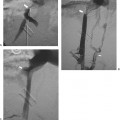
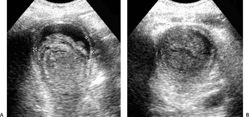
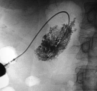
CASE 44 A 72-year-old male in the intensive care unit after coronary artery bypass surgery developed abdominal pain and fever. Physical exam revealed right upper quadrant tenderness and laboratory results revealed leukocytosis. Figure 44-1 (A,B) Transverse sonographic images show gallbladder distention, wall thickening greater than 3 mm, marked intraluminal sludge, and the absence of gallstones. Sonographic evaluation of the right upper quadrant showed gallbladder wall thickening (5 mm), pericholecystic fluid, absence of gallstones, and intraluminal sludge (Fig. 44-1). The gallbladder was distended. Acalculous cholecystitis (AC). General surgery service was consulted and recommended placement of a cholecystostomy tube. The plan was for open cholecystectomy after a few weeks if the patient did not recover despite percutaneous drainage and concomitant antibiotic coverage. Using an Accustick system (Boston Scientific, Natick, Massachusetts) and the Seldinger technique, a 21-gauge needle was advanced into the gallbladder through a small wedge of normal liver parenchyma under direct sonographic guidance. Through the coaxial dilator, a stiff Amplatz wire (Cook, Bloomington, Indiana) was advanced into the gallbladder. Serial fascial dilatation was performed, and an 8-French (F) all-purpose pigtail catheter was placed in the gallbladder lumen (Fig. 44-2). After 5 weeks, the patient was afebrile with a normal white cell count. The catheter was removed without complication, and no further treatment was necessary. Accustick system (Boston Scientific, Natick, Massachusetts) Amplatz wire (Boston Scientific, Natick, Massachusetts) 7 and 9F fascial dilators (Cook, Bloomington, Indiana) 8F multipurpose drainage catheter (Cook, Bloomington, Indiana)
Clinical Presentation
Radiologic Studies
Diagnosis
Treatment
Equipment
Discussion
Background
Stay updated, free articles. Join our Telegram channel

Full access? Get Clinical Tree


