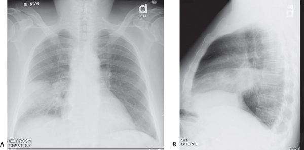
CASE 49 59-year-old man with high fever and cough productive of rusty, blood-streaked sputum PA (Fig. 49.1A) and lateral (Fig. 49.1B) chest X-rays reveal dense, non-segmental homogeneous lobar consolidation that partially silhouettes the right heart border (Fig. 49.1A) and is bordered by the horizontal and oblique fissure (Fig. 49.1B). No associated volume loss. A small ipsilateral pleural effusion blunts the posterior sulcus (Fig. 49.1B). Pneumococcal Pneumonia; Right Middle Lobe • Other Community-Acquired Pneumonias Most pneumococcal infections occur in the winter and early spring. The organism is acquired through person-to-person transmission by inhalation of aerosolized droplets, physical contact, or both. Risk factors for infection include very young or advanced age, chronic heart and/or lung disease, alcoholism and/or cirrhosis, sickle cell anemia, lymphoma, leukemia, multiple myeloma, HIV-AIDS, intravenous drug abuse (IVDA), and prior splenectomy. Fig. 49.1 Pneumococcal pneumonia is caused by the Gram-positive, diplococcal bacterium Streptococcus pneumoniae. This bacterium is responsible for at least half of all cases of community-acquired pneumonia and is also the most common cause of community-acquired pneumonia resulting in hospital admission. Pneumococci colonize the upper respiratory tract of 5–60% of the general population. Pneumococcal pneumonia is the most common bacterial pneumonia to follow influenza pneumonia infection. Additionally, up to 70% of patients with pneumococcal pneumonia have had a recent upper respiratory tract infection (URI). Affected patients often have an abrupt onset of rigors, followed by fever, cough productive of rusty sputum, and dyspnea. Pleuritic chest pain is common, experienced in up to 75% of patients. Leukocytosis is usually present (WBC 15,000–25,000), but severe infection may be associated with white blood cell counts of less than 3,000/mm3. • Metastatic infection (e.g., endocarditis, meningitis, arthritis) • Pericarditis • Rarely empyema • Pneumatoceles; more common in children • Homogeneous, non-segmental, parenchymal consolidation involving one lobe; multi-lobar involvement less common (Figs. 49.1, 49.2A, 49.4) • Air space disease abuts surrounding visceral pleura of fissural surfaces (Figs. 49.1, 49.4) • Predilection for lower lobes or posterior segments of upper lobes (Figs. 49.4, 49.5) • Minimal volume loss (Figs. 49.1, 49.2A
 Clinical Presentation
 Radiologic Findings
 Diagnosis
 Differential Diagnosis
 Discussion
Background
Etiology
Clinical Findings
Complications
Imaging Findings
Chest Radiography
![]()
Stay updated, free articles. Join our Telegram channel


Full access? Get Clinical Tree


