Case 51
Indication: Screening.
History: Thoracotomy at the age of 1 year. Changes in scar texture.
Risk profile: No increased risk.
Age: 45 years.
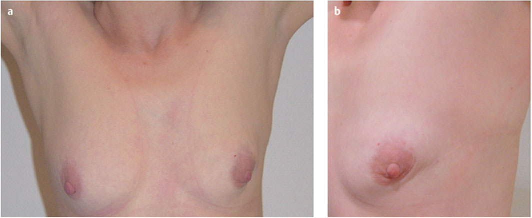
Fig. 51.1 a,b Clinical examination. The patient underwent thoracotomy in infancy for patent ductus arteriosus.
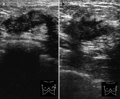
Fig. 51.2a,b Sonography.
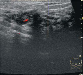
Fig. 51.3 Color-coded Doppler sonography.
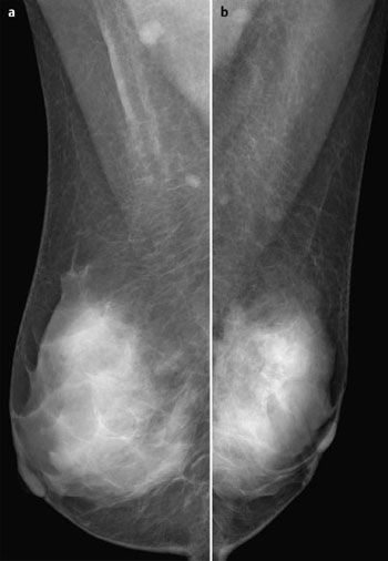
Fig. 51.4a,b Digital mammography, MLO view
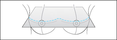
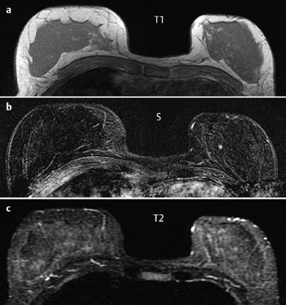
Fig. 51.5a–c Contrast-enhanced MRI of the breasts.
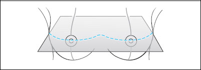
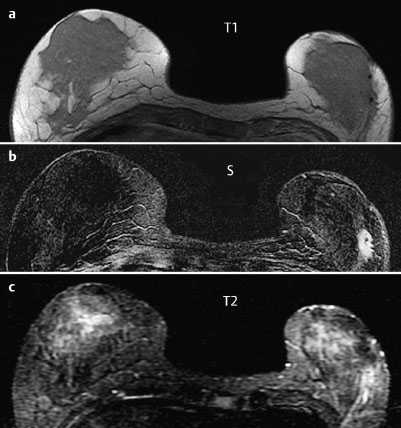
Fig. 51.6a–c Contrast-enhanced MRI of the breasts.
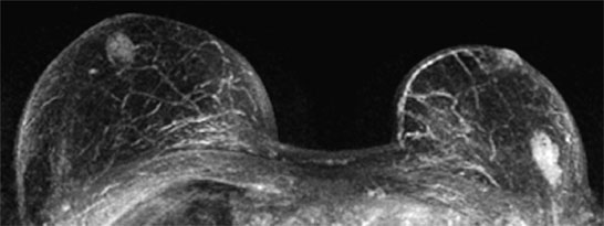
Fig. 51.7 Contrast-enhanced MR mammography. Maximum intensity projection.
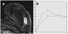
Fig. 51.8a,b Signal-to-time curves.
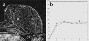
Fig. 51.9a,b Signal-to-time curves.

|
please characterize ultrasound, mammography, and MRI findings.
What is your preliminary diagnosis?
What are your next steps?
|


