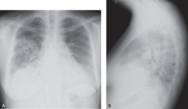
CASE 51 42-year-old woman with long-standing history of poorly controlled diabetes presented to the Emergency Department with purulent productive cough, shortness of breath, and fever PA (Fig. 51.1A) and lateral (Fig. 51.1B) chest X-rays demonstrate poorly defined nodular opacities and patchy areas of consolidation and a small right pleural effusion. Sputum and blood cultures confirmed the diagnosis. Haemophilus influenzae Pneumonia • Other Community-Acquired Bronchopneumonias Fig. 51.1 H. influenzae pulmonary infection is acquired through person-to-person transmission via aerosolized droplets deposited in the nasopharynx. The nasopharynx is colonized in up to 90% of children by 5 years of age. Many patients with COPD are also colonized. The pleomorphic, Gram-negative bacterium Haemophilus influenzae causes Haemophilus pneumonia. Most organisms that colonize the nasopharynx are unencapsulated and non-typeable. Most strains that infect the lung are encapsulated and typeable. Type b is the most common strain responsible for Haemophilus pneumonia (Hib). H. influenzae
 Clinical Presentation
 Radiologic Findings
 Diagnosis
 Differential Diagnosis
 Discussion
Background
Etiology
Clinical Findings
![]()
Stay updated, free articles. Join our Telegram channel


Full access? Get Clinical Tree


