
MRM score | Finding | Points |
Shape | linear | 1 |
Border | ill-defined | 1 |
CM Distribution | homogenous | 0 |
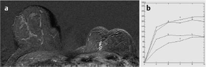
Initial Signal Intensity Increase | strong | 2 |
Post-initial Signal Intensity Character | plateau | 1 |
MRI score (points) |
| 5 |
MRI BI-RADS |
| 4 |
 Differential Diagnostic Considerations
Tumor recurrence, focal mastitis, fat necrosis.
Clinical Findings | right 1 | left 1 |
Ultrasound | right 1 | left 1 |
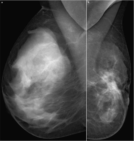
Mammography | right 1 | left 3 |
MR Mammography | right 1 | left 4 |
BI-RADS Total | right 1 | left 4 |
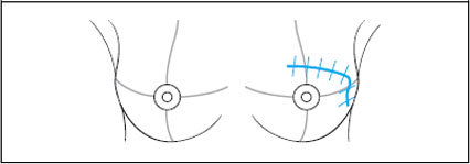
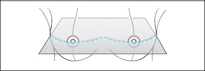
Procedure
MR-guided percutaneous biopsy of the circumscribed enhancement near the chest wall in the left breast.
However, the patient declined this intervention and instead follow-up MR mammography was performed 6 months later.
Follow-up MRI after 6 months
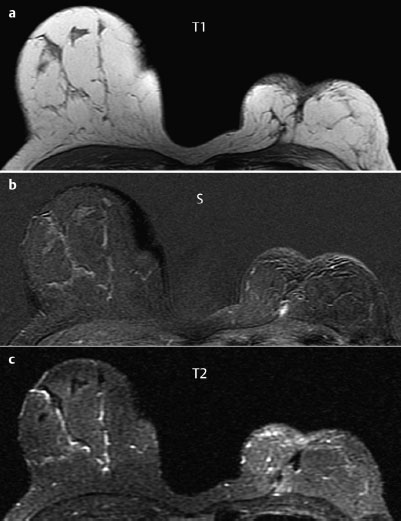
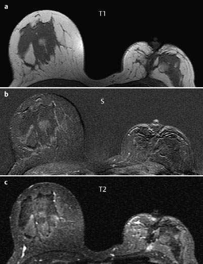
The previously seen linear enhancement in the left breast close to the chest wall was not reproduced to the same extent (Fig. 52.6).
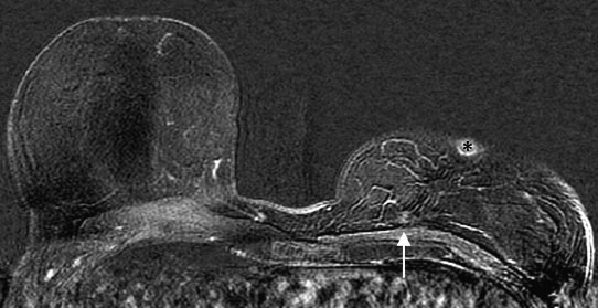
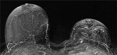
Fig. 52.6 Single-slice subtraction image from the follow-up MRI six months later showing residual contrast uptake (arrow). Enhancement of the nipple (asterisk).
Diagnosis (without histological verification)
Focal inflammation after surgery (breast conservation therapy, intervention after relapse).

Stay updated, free articles. Join our Telegram channel

Full access? Get Clinical Tree


