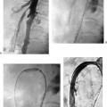
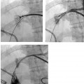
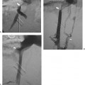
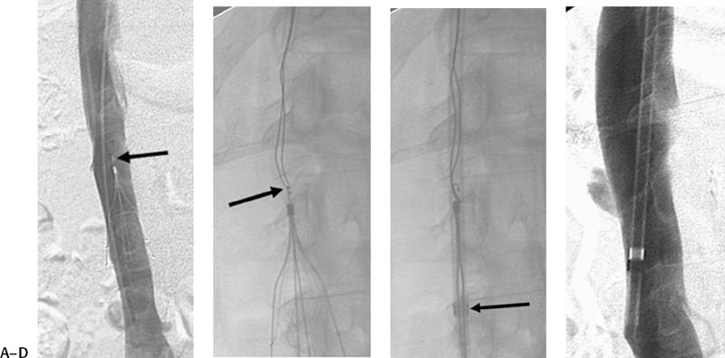
CASE 53 A 34-year-old female with a body mass index of 57 presented for inferior vena cava (IVC) filter placement prior to bariatric surgery. Figure 53-1 A 34-year-old female presented for retrievable IVC filter implantation. A venogram of the IVC was obtained after placement of a Günther Tulip filter below the level of the renal vein without significant tilt. The patient returned for filter retrieval. (A) Initial IVC venogram shows no filling defect within the filter. (B) The hook of the filter was snared. (C) The filter was sheathed by holding the snare still while advancing the sheath. (D) Postretrieval venogram shows an unremarkable IVC. None. Puncture needle or micropuncture kit 5F pigtail and endhole catheters Temporary vena cava filters described above Contrast material The right internal jugular vein was punctured using the Seldinger technique, and the needle was exchanged for a pigtail catheter that was positioned in the IVC. Cavography revealed normal caliber of the IVC without evidence of thrombus or caval variants. Renal vein inflows were identified. The catheter was exchanged over a guidewire for the deployment sheath of the jugular Günther Tulip kit (Cook, Bloomington, Indiana), which was positioned in the infrarenal IVC. In this filter system, a hook is located at the top of the filter. The filter was advanced into the introducer sheath then advanced to the infrarenal IVC level. The sheath was then withdrawn, and the filter was uncovered but still tethered at the hook. The filter was then released by pushing on an external release button that disengages the hook from the introducer system. From the jugular approach, the filter can be repeatedly captured until satisfactory location and alignment are obtained. Six weeks later, after successful gastric bypass surgery and a 60-lb weight loss, the patient returned for filter retrieval. Prior to retrieval, the patient had a negative color duplex study of the lower extremities. Retrieval was performed through a right internal jugular vein access. A guidewire was carefully advanced past the filter under fluoroscopic visualization and over this wire, a 5-French (F) pigtail flush catheter was advanced to the confluence of the iliac vein, and an IVC venogram was performed. This revealed a patent IVC. No clots were detected (Fig. 53-1A). Using the retrieval snare set (Cook, Bloomington, Indiana), the 11F sheath was advanced over an Amplatz wire (Boston Scientific, Natick, Massachusetts) to the top of the filter. The wire and dilator were removed. The snare from the kit was advanced through the sheath, and the hook of the filter was snared (Fig. 53-1B). The filter was sheathed by advancing the sheath forward (Fig. 53-1C). Once in the sheath, the filter was pulled out. A postprocedure IVC venogram showed no evidence of IVC injury (Fig. 53-1D). Pulmonary embolism (PE) remains a major cause of morbidity and mortality and is the most preventable cause of death among hospitalized patients. Anticoagulation is the preferred therapy for deep venous thrombosis (DVT). However, PE may occur despite adequate anticoagulation. Patients with contraindications or complications of anticoagulation may benefit from caval filtration. Numerous studies have documented the safety and efficacy of permanent IVC filters in the prevention of PE in patients with contraindications to anticoagulation or in patients who have failed anticoagulation. However, there are risks associated with long-term implantation of these devices, including symptomatic IVC thrombosis. Strieff, in a compilation of filter data, found the rate of IVC thrombosis to be from 3.6 to 11.2%. Others report a much higher rate of caval thrombosis of up to 50%. Long-term complications also include migration, fracture, DVT, and IVC perforation with or without involvement of adjacent structures.
Clinical Presentation
Radiologic Studies
Treatment
Equipment
IVC Filter Insertion
Follow-up
Retrieval Procedure
Discussion
Background
Noninvasive Imaging Work-up
Treatment Options
Types of Temporary Filters
GÜNTHER TULIP FILTER
Stay updated, free articles. Join our Telegram channel

Full access? Get Clinical Tree


