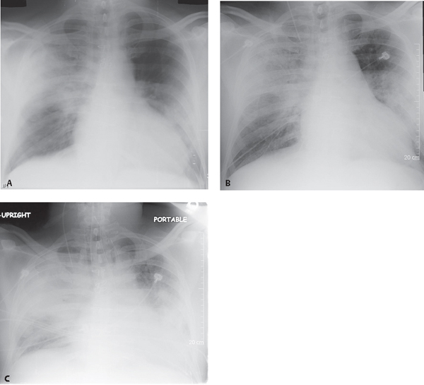
CASE 54 53-year-old man with past CABG on chronic corticosteroids for remote kidney transplant presents with fever, non-productive cough, altered mental status, and diarrhea Baseline AP chest X-ray (Fig. 54.1A) shows patchy, non-segmental heterogeneous right upper lobe and bilateral perihilar opacities. AP chest X-ray two days later (Fig. 54.1B) demonstrates progression of disease, although confined to the same regions of lung. Follow-up chest X-ray two days later (Fig. 54.1C) reveals rapid progression of disease. Air space consolidation now replaces most of the right upper and left lower lobes. The left perihilar disease has progressed and extends into the upper lobe. Hypoxia necessitated intubation. Fig. 54.1 Legionella Pneumonia • Other Community-Acquired Bronchopneumonias Legionella was so named after a July 1976 outbreak of an unknown illness in Philadelphia at the American Legion Convention, during which 221 persons became ill and 34 died. On January 18, 1977, the unknown bacterium was identified and subsequently named Legionella. Since then, at least 50 species of Legionella have been identified. Pulmonary infection with Legionella
 Clinical Presentation
 Radiologic Findings

 Diagnosis
 Differential Diagnosis
 Discussion
Background
![]()
Stay updated, free articles. Join our Telegram channel

Full access? Get Clinical Tree


