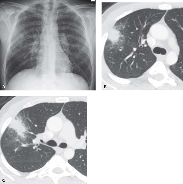
CASE 55 46-year-old man with chest pain, cough, and night sweats for several weeks PA chest radiograph (Fig. 55.1A) demonstrates patchy non-segmental right mid-lung air space disease, bilateral hilar lymphadenopathy, and poor visualization of the right posterolateral fifth rib. Unenhanced chest CT (lung window) (Figs. 55.1B, 55.1C) demonstrates a peripheral right upper lobe mass-like consolidation with a surrounding halo of ground glass opacity. Pulmonary Nocardiosis Fig. 55.1 • Other Atypical Pulmonary Infection (including tuberculosis and fungal infection) • Septic Embolus • Primary Lung Cancer • Pulmonary Vasculitis
 Clinical Presentation
 Radiologic Findings
 Diagnosis

 Differential Diagnosis
 Discussion
Background
Stay updated, free articles. Join our Telegram channel

Full access? Get Clinical Tree


