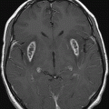
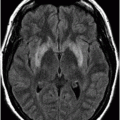
Axial FLAIR through the level of the lower pons.
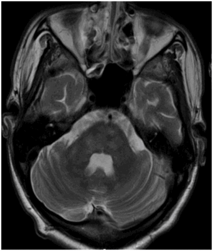
Axial T2WI through the level of the lower pons.
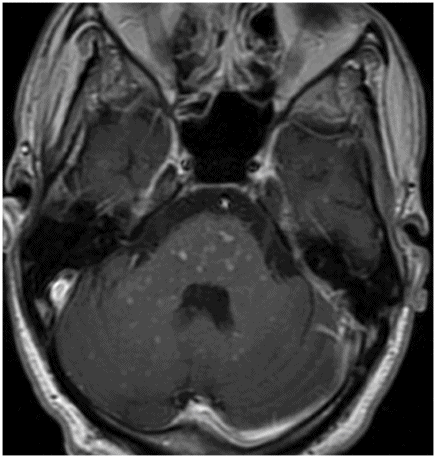
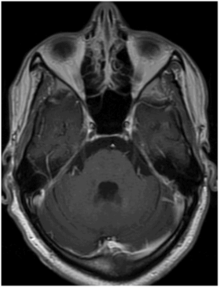
Axial postcontrast T1WI image through the pons.
CLIPPERS
Primary Diagnosis
CLIPPERS
Differential Diagnoses
Lymphomatoid granulomatosis
Vasculitis (primary CNS vasculitis or CNS manifestation of systemic vasculitides)
Neurosarcoidosis
Angiocentric lymphoma
Imaging Findings
Fig. 56.1: Axial FLAIR and Fig. 56.2: Axial T2WI images through the level of the lower pons demonstrated multiple, subtle punctate areas of increased T2 signal centered in the pons and in the middle cerebellar peduncles. Fig. 56.3: Axial postcontrast T1WI image through the same level demonstrated punctate areas of enhancement in the respective areas of T2 hyperintensities. Fig. 56.4: Axial postcontrast T1WI after a high dose of contrast through the same level demonstrated complete resolution of the enhancing foci.
Discussion
The presence of subacute-onset cerebellar and brainstem symptoms, responsiveness to steroids, and signature imaging findings – punctate foci of enhancement peppering in the brainstem and cerebellum – are consistent with a diagnosis of chronic lymphocytic inflammation with pontine perivascular enhancement responsive to steroids (CLIPPERS).
Perivascular enhancement can be seen in lymphomatoid granulomatosis, sarcoidosis, vasculitis (including primary CNS angiitis and CNS manifestation of systemic vasculitides), and lymphoma. Predominantly involving the lungs, lymphomatoid granulomatosis is a rare Epstein-Barr virus-associated, angiodestructive, lymphoproliferative disorder that seldom involves the CNS. Diffuse punctate enhancement that may have perivascular distribution is seen on imaging. In the case of brain involvement, punctate enhancement involves the entire brain with no predilection for the pons or cerebellum. Clinical manifestation of CLIPPERS clearly differs from that of previously mentioned differential diagnoses.
Dural manifestations of neurosarcoidosis are commonly seen in the lepto- and pachymeninges. Perivascular enhancement can also be seen; however, it is characteristically limited to the basal ganglia, hypothalamus, and inferior frontal lobe. In addition, tumor-like involvement of the brain parenchyma is a known manifestation of neurosarcoidosis. Sarcoid granulomas are typically hypointense on T2WI images that demonstrate intense contrast enhancement. Primary CNS angiitis does not have any associated imaging abnormalities. Focal cortical or subcortical infarcts are common manifestations with or without meningeal enhancement. Central nervous system lymphoma typically demonstrates a homogeneously enhancing mass with diffusion restriction. Perivascular spread is a known manifestation of lymphoma; however, the enhancement is linear along the perivascular space, rather than punctate. Unlike CLIPPERS, isolated pontine and cerebellar involvement has not been described in perivascular lymphoma spread.
Bickerstaff brainstem encephalitis (BBE) and neuro-Behçet disease (NBD) have a predilection for the posterior fossa structures. Bickerstaff brainstem encephalitis is usually preceded by systemic illness. Affected individuals commonly present with a decreased level of consciousness and brainstem and cerebellar signs. Abnormalities on MRI can be seen in up to 31% of patients and include non-enhancing T2 signal abnormality. Central nervous system involvement in Behçet disease is rare and typically occurs three to five years after disease onset. Characteristic NBD imaging anomalies include asymmetric T2 abnormalities along the white matter tract involving 1) the mesodiencephalic junction (46%); 2) pons (40%); 3) hypothalamus and thalamus (23%); and 4) basal ganglia (18%). Although enhancement is uncommon in NBD, ring-like, patchy, and nodular, non-confluent enhancement patterns without any tropism for perivascular spaces in the cerebellum or pons have been described in patients with NBD.
CLIPPERS is a definable, treatable, posterior fossa-predominant inflammatory disorder of unknown etiology. It has a characteristic clinical presentation: CNS lesions that are typical in their distribution, histology, and imaging morphology. Manifestation of typical clinical presentation is dependent upon the areas of brain involvement and includes sensory abnormalities, diplopia, ataxia, and dysarthria. Symptom onset is typically subacute. Diplopia and ataxia are the most common presenting symptoms. Ataxia, diplopia, ataxic dysarthria, altered facial sensation, dizziness, nausea, tremor, and pseudobulbar affect appear in varying combinations during disease progression. The age of presentation varies between 16 and 86 years of age, with no sex predilection.
The distinguishing imaging features of CLIPPERS include the presence of punctate and curvilinear enhancement peppering the pons with superior extension to the midbrain, inferior extension to the medulla, and posterior extension to the middle cerebellar peduncles with or without cerebellar involvement. Most case series report varied cerebellum involvement. As a result, a revised name, chronic lymphocytic inflammation with pontocerebellar perivascular enhancement responsive to steroids has been suggested. Typically, the densest population of enhancing foci is centered at the pons with the number and size of enhancing foci decreasing as the distance from the pons increases. Most lesions are typically less than 5 mm in diameter; few may be 1 cm or more in diameter. Tiny ring enhancement has also been described in CLIPPERS. Rarely, lesions can be seen in the basal ganglia or the spinal cord; however, even in these cases the densest population remains in the pons, the characteristic imaging abnormality. Involved areas appear as patchy or confluent areas of hyperintensity on T2WI images, including FLAIR, rather than as punctate hyperintensity. Mass effect is typically absent. There may be a variable degree of patchy diffusion restriction.
Typical histopathology findings include marked perivascular CD3-predominent lymphocytic infiltration in the white matter, in addition to presence of CD20-positive B cells, and CD68-positive histiocytes without any evidence of demyelination or granuloma formation. Inflammation of the vessel wall, as characteristically seen in vasculitis, is typically absent in CLIPPERS.
As the acronym suggests, CLIPPERS responds to steroids. Rapid tapering of steroid dose may cause relapse. Relapse-free intervals increase with slower steroid tapering. Low-dose methotrexate has also been recommended to increase length of relapse-free intervals. Enhancing foci gradually disappear with successful treatment and diffusion restriction reverses. Gradually, atrophy of the involved areas is readily appreciated in the cerebellum.
Stay updated, free articles. Join our Telegram channel

Full access? Get Clinical Tree


