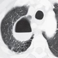
CASE 56 39-year-old man presents with fever, cough, sputum production, and left-sided chest pain PA chest radiograph (Fig. 56.1A) shows an ill-defined left upper lobe perihilar mass-like opacity without air bronchograms. Contrast-enhanced chest CT (mediastinal window) (Fig. 56.1B) reveals a mass-like region of consolidation in the medial anterior segment of the left upper lobe that invades the adjacent anterior mediastinum and left anterior chest wall. Note the heterogeneous attenuation infiltrative process producing asymmetric thickening of the left pectoralis muscles. Pulmonary Actinomycosis with Chest Wall Invasion • Atypical Infections (including nocardiosis, tuberculosis, and fungal infections) • Primary Lung Cancer
 Clinical Presentation
 Radiologic Findings
 Diagnosis
 Differential Diagnosis
 Discussion
Background
Stay updated, free articles. Join our Telegram channel

Full access? Get Clinical Tree


