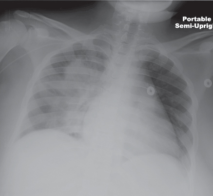
CASE 57 Morbidly obese woman hospitalized for management of her diabetes who developed fever, productive cough, and dyspnea during her hospitalization AP (Fig. 57.1) chest radiograph reveals diffuse right perihilar nonsegmental air space disease without cavitation, pleural effusion, or lymphadenopathy. Subsequent bronchoscopy confirmed the diagnosis. Acinetobacter Hospital-Acquired Pneumonia • Nosocomial Hospital-Acquired Pneumonia of Other Etiologies • Aspiration Pneumonia • Community-Acquired Pneumonias Acinetobacter species are Gram-negative, nonmotile, aerobic coccobacillary organisms. However, they can be Gram-variable and occasionally Gram-positive on initial stains. The morphologic appearance is dependent on the growth phase with a rod-shaped appearance during rapid growth but a coccobacillary appearance during the stationary phase. Acinetobacter species are oxidase-negative, which differentiates this species from other Gram-negative organisms, such as Pseudomonas, Neisseria, and Moraxella. Fig. 57.1 (Image courtesy of Kristin Miller, MD, VCU Medical Center, Richmond, Virginia.) Acinetobacter grow easily in nature and are most commonly found in the soil and water. However, Acinetobacter has also been isolated from food, ventilator and suctioning equipment, infusion pumps, sinks, pillows, bed mattresses and railings, tap water, humidifiers, soap dispensers, etc. Such colonization is extremely problematic, as Acinetobacter survives an average of 20 days and can survive for as long as four months on hospital surfaces. Furthermore, up to 40% of healthy adults have skin colonization, with even higher rates among hospital employees and patients. The respiratory tract is frequently colonized in humans and is the most common site of infection. Acinetobacter is primarily a colonizer of the hospital environment and of the ICU in particular and is an important cause of hospital-acquired (HAP) and ventilator-acquired (VAP) pneumonia. Acinetobacter
 Clinical Presentation
 Radiologic Findings
 Diagnosis
 Differential Diagnosis
 Discussion
Background

Etiology
Clinical Findings
![]()
Stay updated, free articles. Join our Telegram channel

Full access? Get Clinical Tree


