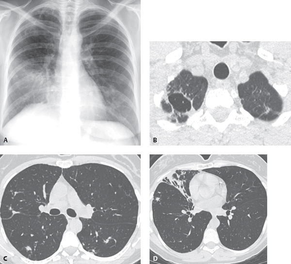
CASE 60 40-year-old woman with cough and weight loss PA chest radiograph (Fig. 60.1A) demonstrates middle lobe air space disease, a right apical thin-walled cavity, and multifocal bilateral lung nodules. Unenhanced thin-section chest CT (lung window) (Figs. 60.1B, 60.1C, 60.1D) demonstrates a thin-walled right apical lung cavity with surrounding architectural distortion (Fig. 60.1B), middle lobe atelectasis with intrinsic bronchiectasis (Fig. 60.1D), and scattered lung nodules with irregular borders (Fig. 60.1C). Fig. 60.1 Mycobacterium avium-intracellulare Pulmonary Infection • Other Pulmonary Infections (including tuberculosis, other non-tuberculous mycobacteria, and aspergillus) Mycobacteria other than M. tuberculosis cause pulmonary and systemic infection in immunocompetent and immunocompromised individuals. These organisms occur ubiquitously in the environment (soil, water, plants, and animals). Non-tuberculous mycobacterial infections are produced by a variety of mycobacteria, including M. aviumintracellulare (MAI), M. avium complex (MAC), M. kansasii, M. fortuitum, M. cheloneae, M. abscessus, and M. xenopi. It is thought that infection occurs from environmental exposures as there are no documented cases of person-to-person transmission. Infection may occur through inhalation or via the gastrointestinal tract. Nontuberculous mycobacterial pulmonary infection may exhibit various imaging manifestations depending on the patient population affected. The classic form of infection typically affects elderly Caucasian men with underlying chronic lung disease and is indistinguishable from pulmonary tuberculosis. The bronchiectatic form of infection (non-classic
 Clinical Presentation
 Radiologic Findings
 Diagnosis
 Differential Diagnosis
 Discussion
Background
Etiology
Clinical Findings
![]()
Stay updated, free articles. Join our Telegram channel


Full access? Get Clinical Tree


