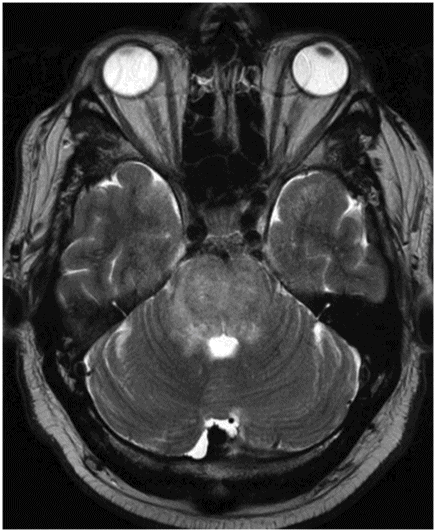
Axial FLAIR image through the level of the pons.
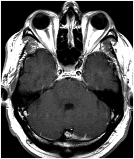
Axial T2 image through the same level.
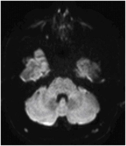
DWI image through the same level.
Central Variant of Posterior Reversible Encephalopathy Syndrome (CV-PRES)
Primary Diagnosis
Central variant of posterior reversible encephalopathy syndrome (CV-PRES)
Differential Diagnoses
Rhombencephalitis
Brainstem gliomas
Osmotic demyelination syndrome
Tumefactive demyelination
Imaging Findings
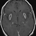
Fig. 62.1: Axial FLAIR image and Fig. 62.2: Axial T2 image through the level of the pons. Images demonstrated diffuse swelling of the pons associated with T2 hyperintensity that caused minimal mass effect on the fourth ventricle floor. Fig. 62.3: DWI image through the same level does not demonstrate any diffusion restriction or facilitation. Fig. 62.4: Axial postcontrast image through the same level does not demonstrate any abnormal enhancement. On follow-up imaging, all the abnormalities were resolved.
Discussion
This clinical presentation is classic for a hypertensive emergency. Central variant of posterior reversible encephalopathy syndrome (CV-PRES) is a known variant of PRES. It involves the central brain structures such as basal ganglia, thalami, periventricular white matter, and brainstem instead of the classic posterior predominant supratentorial brain involvement seen in typical PRES. In the given clinical context, this imaging manifestation is diagnostic of CV-PRES.
Rhombencephalitis (RE) or brainstem encephalitis is a syndrome of multiple causes and outcomes. Although RE can have an identical imaging appearance, it has a more subacute onset. Most common infectious causes of rhombencephalitis stem from infectious etiologies such as Listeria monocytogenes, enterovirus, or herpes simplex virus type 1. In addition to infectious causes, paraneoplastic syndrome and autoimmune diseases such as Behçet disease, may also present as rhombencephalitis. Although diffuse pontine glioma can have a similar imaging appearance, its clinical presentation is different. Osmotic demyelination syndrome (ODS) typically presents acutely after rapid correction of hyponatremia. Acute demyelination in ODS involves the central pontine white matter, and spares the peripheral pontine structures, rather than involving the global pontine. Rarely, tumefactive demyelination involves the brainstem and in such cases, presentation is different. Typically, acute tumefactive lesion presents with enhancement, which is absent in this patient.
Posterior reversible encephalopathy syndrome is a clinicoradiologic diagnosis with typical predisposing factors and typical imaging appearances. It typically occurs in two different clinical contexts: 1) as a manifestation of hypertensive urgency or emergency, and 2) as a complication of CNS toxicity from different drug therapies such as immunomodulator, chemotherapeutic, or monoclonal antibody therapy.
The pathophysiologic manifestation of PRES is still not fully understood. In patients with hypertension, it is thought to arise from failed autoregulation, resulting in breakthrough brain edema versus severe autoregulatory vasoconstriction induced by hypertension, resulting in brain ischemia and edema. T-cell activation with inflammatory cytokine production or endothelial activation (with or without leukocyte trafficking) are common underlying biologic processes manifested in PRES.
Clinical presentation of PRES is variable, and depends largely upon the areas of brain involvement and disease severity. Common symptoms include headache, vision change, extremity weakness, nausea, and altered mental status. Symptoms may develop acutely or sometimes over several days. In severe widespread brain involvement, generalized seizures and coma may develop.
The typical imaging manifestations of PRES include abnormal FLAIR signal in the cortical and subcortical white matter of the occipital lobes and posterior parietal lobe. Involvement can be symmetric or asymmetric. These findings are typically reversible with treatment of hypertension or withdrawal of the offending drug. Diffusion restriction, hemorrhage, and enhancement are uncommon imaging manifestations and usually signify poorer prognosis. In severe cases, the deep brain structures such as basal ganglia and thalami can also be involved, in addition to the typical cortical and subcortical white matter involvement.
Central variant of PRES is a known variant of PRES and accounts for approximately 10–20% of all PRES cases. In this variant, geographic distribution of PRES is different from the typical PRES. The deeper brain structures such as basal ganglia, thalami, periventricular white matter, and brainstem structures are involved. In addition to the typical predisposing factors, systemic lupus erythematosus and other autoimmune diseases can also present as CV-PRES.
If the clinical presentation of the patient is not typical for PRES and in the absence of specific predisposing factors, the diagnosis of CV-PRES can be challenging as other diseases as discussed above can have identical imaging presentations.


Stay updated, free articles. Join our Telegram channel

Full access? Get Clinical Tree


