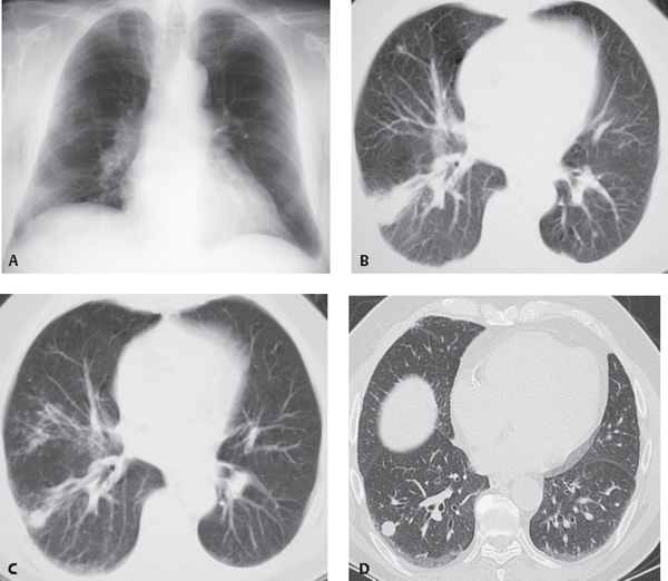
CASE 62 78-year-old man from Arizona with cough, myalgias, and headache PA chest radiograph (Fig. 62.1A) demonstrates ill-defined nodular opacities in the right middle and lower lung zones. Unenhanced chest CT (lung window) (Fig. 62.1B) demonstrates a wedge-shaped right lower lobe consolidation and bilateral small pulmonary nodules. Unenhanced chest CT (lung window) (Fig. 62.1C) obtained six months after presentation demonstrates improvement of the right lower lobe consolidation with a residual pulmonary nodule as well as multiple right middle lobe centrilobular nodules. HRCT (Fig. 62.1D) obtained one year after presentation demonstrates a well-defined right lower lobe solitary pulmonary nodule in the region of the former right lower lobe consolidation. Coccidioidomycosis Fig. 62.1 • Other Fungal Infection • Tuberculosis Coccidioidomycosis is a highly infectious fungal disease endemic to the southwestern United States, northern and central Mexico, and Central and South America. It is estimated that there are 100,000 new cases of infection each year in the United States.
 Clinical Presentation
 Radiologic Findings
 Diagnosis

 Differential Diagnosis
 Discussion
Background
Etiology
Stay updated, free articles. Join our Telegram channel

Full access? Get Clinical Tree


