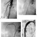
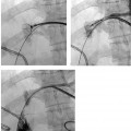
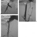
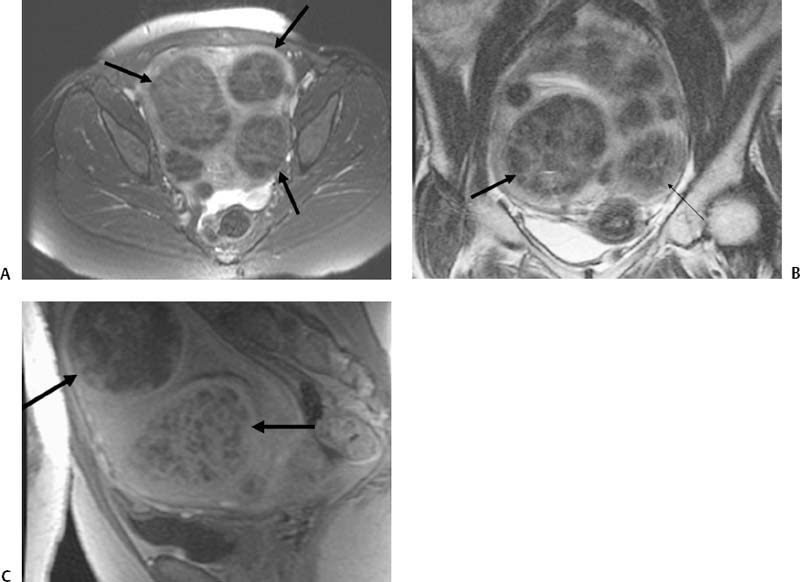
CASE 62 A 44-year-old female presented to us with pelvic pain and menorrhagia. Figure 62-1 A 44-year-old female with pelvic pain and menorrhagia. MRI study was obtained. (A) Axial T1-weighted, (B) coronal T1-weighted, and (C) sagittal T1-weighted post-gadolinium images show an enlarged uterus containing several enhancing masses (arrows), typical for fibroids. There was significant mass effect on the adjacent colon and bladder. Pre- and postgadolinium contrast-enhanced MRI studies were performed. MRI showed the enlarged uterus containing multiple enhancing masses with mass-effect on the colon and bladder (Fig. 62-1). Uterine fibroids. 5F pigtail catheter (AngioDynamics, Queensbury, New York) to perform abdominal aortogram and pelvic arteriogram 5F angled glide catheter (Terumo, Somerset, New Jersey) to select the internal iliac artery and to perform iliac arteriogram Precurved catheters with tapered tip such as the Ann Roberts (Boston Scientific, Natick, Massachusetts) could also be used. If the uterine artery is of sufficient size, its selection can be performed using the same glide catheter, and embolization can be performed using the same catheter. However, if the uterine artery is small, a microcatheter should be used, in the coaxial technique, to select the uterine artery, to perform a uterine arteriogram, and to embolize. Microcatheters commonly used include Tracker 325 (Boston Scientific, Natick, Massachusetts), Renegade (Boston Scientific, Natick, Massachusetts), and Progreat (Terumo, Somerset, New Jersey). These catheters come with their own wires, but other wires could be used. Four agents are available for fibroid embolization: Polyvinyl alcohol particles (Boston Scientific, Natick, Massachusetts) Spherical polyvinyl alcohol particles (Boston Scientific, Natick, Massachusetts) Bead Block (compressible polyvinyl alcoholmicrospheres; Terumo, Somerset, New Jersey) Embospheres (Biosphere Medical, Rockland, Massachusetts) Currently, there is controversy regarding which agent is best for UAE as well as the end point of embolization for particular agents. Use of Gelfoam (Upjohn and Pharmacia, Kalamazoo, MI) for this purpose is not widespread but has been reported. The patient was admitted to the interventional radiology service (Table 62-1). Preprocedural antibiotics were administered, and a Foley catheter was inserted into the bladder. A 5-French (F) catheter was inserted into the common femoral artery, and a pelvic arteriogram was obtained (Fig. 62-2) with a 5F pigtail catheter. The pigtail catheter was exchanged over a guidewire for a 5F glide catheter (Boston Scientific, Natick, Massachusetts), which was used to selectively catheterize the left uterine artery. Particles were infused (Embospheres, 500 to 700 microns, followed by 700 to 900 microns; Biosphere Medical, Rockland, Massachusetts) to occlude blood flow. The catheter was then used to form a Waltman’s loop in the abdominal aorta and then to selectively catheterize the ipsilateral right uterine artery. Particle embolization was performed to occlude blood flow. The catheter was then removed, and hemostasis was achieved. The patient was admitted overnight with a patient-controlled analgesic pump. She was converted to oral medications on the subsequent day and discharged when pain control was adequate (Table 62-2).
Clinical Presentation
Radiologic Studies
Magnetic Resonance Imaging (MRI)
Diagnosis
Treatment
Equipment
ANGIOGRAPHIC CATHETERS
EMBOLIZATION AGENTS
Uterine Artery Embolization (UAE)
| Interventional radiology consultation Review patient’s history and imaging to ensure procedure is warranted. Discuss risks, benefits, known results, and alternatives to UFE. Establish rapport with patient. Evaluation by gynecologist Endometrial biopsy to exclude endometrial cancer Exclude other causes of symptoms. Ensure continued follow-up by gynecologist after procedure. Pelvic ultrasound or MRI Evaluate for fibroid burden. Look for concurrent disease process or other explanation for symptoms. |
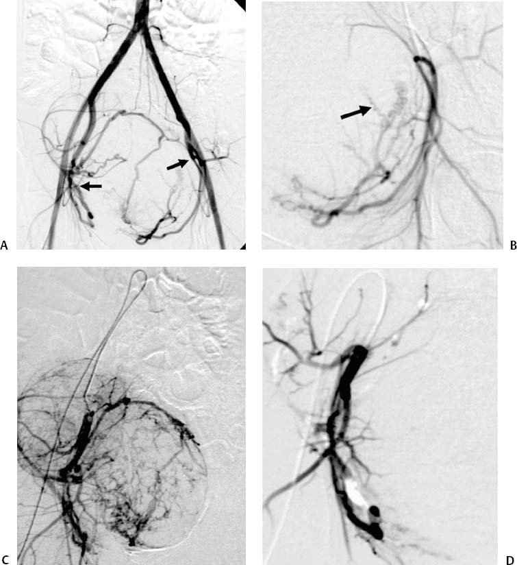
Figure 62-2 UAE. (A
Stay updated, free articles. Join our Telegram channel

Full access? Get Clinical Tree


