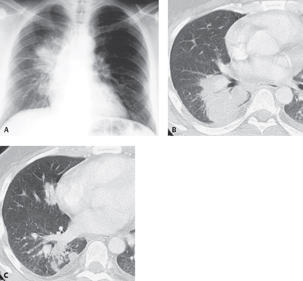
CASE 63 39-year-old man with cough, fever, chills, and skin lesions PA chest radiograph (Fig. 63.1A) demonstrates a large mass-like consolidation in the right perihilar region affecting the right upper and lower lobes. Coned-down contrast-enhanced chest CT (lung window) (Figs. 63.1B, 63.1C) demonstrates a mass-like consolidation in the right lower lobe with central air bronchograms (Fig. 63.1B) and multifocal pulmonary nodules in the right lower and middle lobes (Fig. 63.1C). Note trace bilateral pleural effusions. Blastomycosis Fig. 63.1 • Primary Lung Cancer • Lung Abscess • Round Pneumonia North American blastomycosis is a fungal infection endemic to the Mississippi and Ohio River valleys, the midwestern United States, the Canadian provinces near the Great Lakes, and the Saint Lawrence River valley, as well as other regions of the Americas, Europe, and Asia. The fungus grows in nitrogen-rich soil in wooded areas near streams, rivers, and lakes. Blastomycosis is caused by Blastomyces dermatitidis, a fungus acquired by inhalation of infected soil. Many outbreaks occur near recreational water.
 Clinical Presentation
 Radiologic Findings
 Diagnosis
 Differential Diagnosis
 Discussion
Background
Etiology
Clinical Findings
Related posts:


Stay updated, free articles. Join our Telegram channel

Full access? Get Clinical Tree


