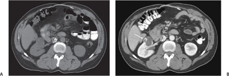
CASE 64 A 62-year-old patient complains of right flank pain. Fig. 64.1 (A) Noncontrast CT image shows an inferior vena cava filter in place. There is an exophytic low-attenuating lesion arising from the right kidney, with thickening seen along the right lateral wall. (B) Following the administration of contrast, there is an enhancing nodule (arrow) seen within the lesion. In addition, there are ill-defined septations and thickening of the left lateral wall. Noncontrast computed tomography (CT) image shows an inferior vena cava filter in place. There is an exophytic low-attenuating lesion arising from the right kidney, with thickening seen along the right lateral wall (Fig. 64.1A). Following the administration of contrast, there is an enhancing nodule seen within the lesion (Fig. 64.1B). In addition, there are ill-defined septations and thickening of the left lateral wall. Cystic renal cell carcinoma (RCC) RCCs account for 85% of all renal malignancies. They are usually solid, but up to 20% can be cystic.
Clinical Presentation
Radiologic Findings
Diagnosis
Differential Diagnosis
Discussion
Background
Related posts:


Stay updated, free articles. Join our Telegram channel

Full access? Get Clinical Tree


