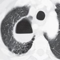
CASE 65 42-year-old debilitated woman with advanced AIDS, cryptococcal meningitis, minimally productive cough, and fever. PA (Fig. 65.1A) and lateral (Fig. 65.1B) chest radiographs demonstrate subtle areas of air space disease in the left upper, right middle, and left lower lobes. No pleural effusion or lymphadenopathy is present. Chest CT (lung window) (Figs. 65.1C, 65.1D, 65.1E) confirms the presence of ground glass and nodular opacities in the left upper lobe, lingula, right middle and lower lobes, and frank left lower lobe consolidation with associated bronchial dilatation. Cryptococcal Pneumonia • Pneumocystis jiroveci Pneumonia • Mycobacterium tuberculosis • Histoplasma capsulatum • Blastomyces dermatitidis • Other Fungal and Atypical Bacterial Pneumonias The genus Cryptococcus contains more than 50 species; however, only C. neoformans and Cryptococcus gattii are considered human pathogens. Each species has five serotypes. C. neoformans is an encapsulated yeast found worldwide and is the most common species in the United States and other temperate climates. C. neoformans
 Clinical Presentation
 Radiologic Findings
 Diagnosis
 Differential Diagnosis
 Discussion
Background
![]()
Stay updated, free articles. Join our Telegram channel

Full access? Get Clinical Tree


