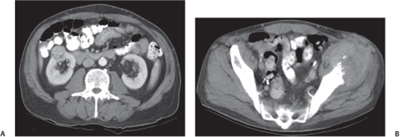
CASE 66 A 67-year-old man presents with hematuria. Fig. 66.1 (A) Axial contrast-enhanced CT image shows a well-defined, exophytic, intensely enhancing solid lesion from the anterior cortex of the right kidney. (B) CT image of the pelvis in the same patient shows a large, round, heterogeneously enhancing soft tissue mass lesion in the anterosuperior iliac spine on the left side, causing bone destruction. Axial contrast-enhanced computed tomography (CT) image shows a well-defined, exophytic, intensely enhancing solid lesion arising from the anterior cortex of the right kidney. There are associated bony metastases in the left ilium (Fig. 66.1). Renal cell carcinoma (RCC) RCC is the most common lethal urologic malignancy. It accounts for 95% of cases of renal tumors. With the growing popularity of imaging studies, the prevalence of RCC has increased, with more than half of the cases being detected coincidentally on the imaging studies done for nonrenal indications. Clear cell is the most common and most aggressive histologic type of RCC. RCC is cystic in 10 to 15% of the total cases of RCC. Most patients are asymptomatic, with the renal lesion detected incidentally on radiologic examination done for other causes. Presenting clinical features can be hematuria, pain in the abdomen or flanks, abdominal mass, weight loss, anorexia, night sweats, fever, or symptoms from distant meta-static disease. Bilateral RCCs can be seen in von Hippel-Lindau disease, tuberous sclerosis, familial RCCs, and acquired renal cystic disease.
Clinical Presentation

Radiologic Findings
Diagnosis
Differential Diagnosis
Discussion
Background
Clinical Findings
Complications
Etiology
Stay updated, free articles. Join our Telegram channel

Full access? Get Clinical Tree


