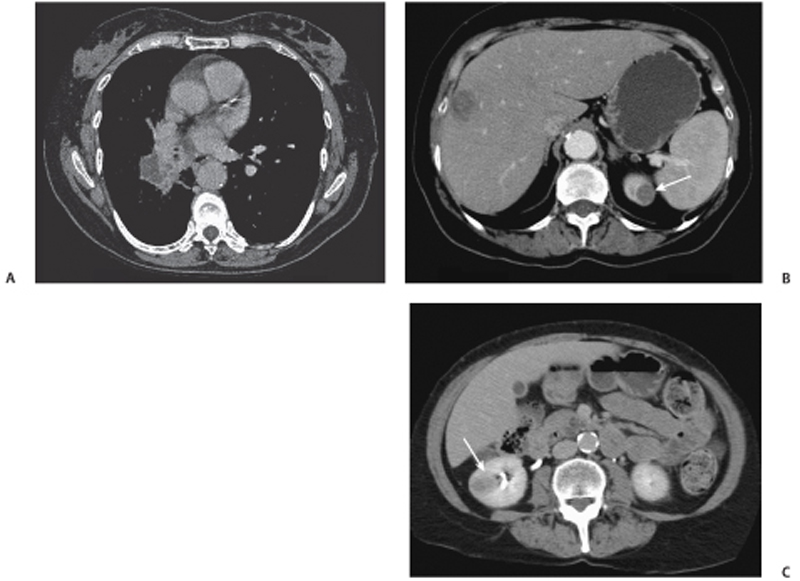
CASE 67 A 59-year-old woman with a known primary history of adenocarcinoma of the lung presents with flank pain. Fig. 67.1 (A) Contrast-enhanced image in a 59-year-old patient shows the presence of an enhancing right hilar adenocarcinoma. (B) A more inferior abdominal axial image shows the presence of a low-density metastatic liver lesion along with a low-density left renal lesion (arrow). (C) An additional low-density lesion is seen in the right kidney (arrow). Axial computed tomography (CT) image of the chest shows the patient’s known primary right lung cancer (Fig. 67.1A). Abdominal images show hypoattenuating hepatic metastases as well as bilateral low-density renal lesions (Fig. 67.1B,C). Renal metastases from primary lung adenocarcinoma
Clinical Presentation
Radiologic Findings
Diagnosis
Differential Diagnosis
Discussion
Related posts:


Stay updated, free articles. Join our Telegram channel

Full access? Get Clinical Tree


