Case 69(Continuation of Case 68)
Indication: Follow-up of findings categorized BI-RADS 3 in the left breast.
History: Unremarkable.
Risk profile: No increased risk.
Age: 77 years.
Clinical Findings
Normal.
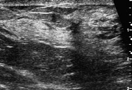
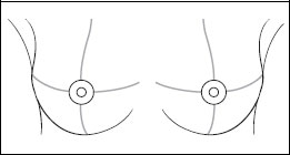
Fig. 69.1 a,b Ultrasound between the outer quadrants of the left breast.
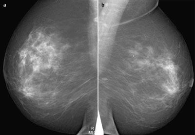
Fig. 69.2 a,b Digital mammography, MLO view.
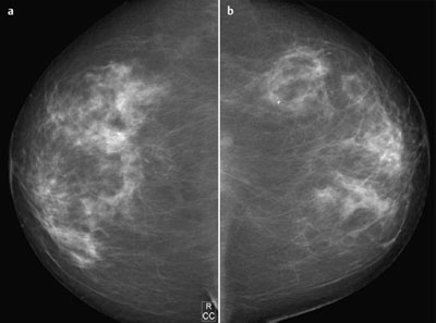
Fig. 69.3 a,b Digital mammography, CC view.
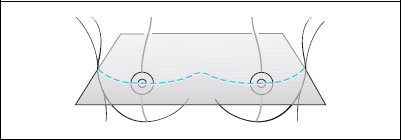
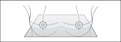
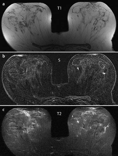
Fig. 69.4 a-c Contrast-enhanced MRI of the breasts.
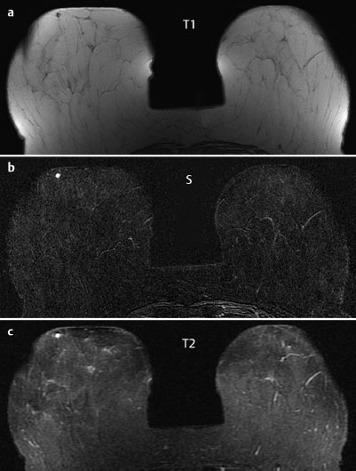
Fig. 69.5 a-c Contrast-enhanced MRI of the breasts.
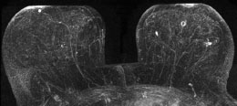
Fig. 69.6 Contrast-enhanced MR mammography. Maximum intensity projection.
| Please characterize ultrasound, mammography, and MRI findings. What is your preliminary diagnosis? What are your next steps? |

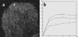
Fig. 69.7 a,b Signal-to-time curves, right breast.
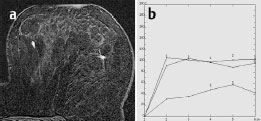
Fig. 69.8 a,b Signal-to-time curves, left outer quadrants.
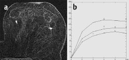
Fig. 69.9 a,b Signal-to-time curves, left inner quadrants.
This case shows the follow-up after 6 months of a lesion depicted in sonography and MRI between the outer quadrants of the left breast (Case 68).
Ultrasound
Again, a hypoechoic, irregular lesion (diameter 4 mm) was seen between the outer quadrants of the left breast. Sonography depicted this lesion only in one plane. A slight increase in possible spiculations was seen. There was also distal shadowing. There were no echogenic margins and there was no architectural distortion. A hypoechoic lesion between the inner quadrants of the left breast (diameter 5 mm) and a cyst (diameter 1 cm) between the lower quadrants of the left breast were visible (these findings were unchanged since the previous examination). US BI-RADS left 4.
Mammography
Inhomogeneously dense parenchyma, ACR type 3. Particularly between the outer quadrants of the left breast, there were still no signs of suspicious lesions. BI-RADS right 1/left 1. PGMI: CC view G (medial skin fold); MLO view M (angle of right pectoralis muscle, left nipple, inframammary fold).
MR Mammography
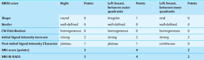
There was an irregular, well-defined lesion between the outer quadrants of the left breast (diameter 5 mm) with homogeneous contrast uptake, an initial signal increase of 110%, and a postinitial plateau as well as reduced signal in T2-weighted imaging. There was also a well-defined lesion measuring 5 mm between the inner quadrants of the left breast with homogeneous enhancement, an initial signal increase of 130%, and a postinitial continuing signal increase as well as increased signal in T2-weighted imaging. Also, unchanged since the previous examination, MRI depicted a well-defined lesion (diameter 4 mm) in the lower outer quadrant of the right breast with an initial signal increase of 130% and a post-initial plateau as well as increased signal in T2-weighted imaging. In maximum intensity projection there was a ring enhancement behind the left nipple with unremarkable signal curve and increased signal in T2-weighted imaging (no quantification, complicated cyst).
MRI Artifact Category: 1
MRI Density Type: 2
 Differential Diagnosis
Left lateral: Adenosis, carcinoma.
Left medial: Fibroadenoma, adenoma, papilloma.
Left behind nipple: Complicated cyst.
Right: Fibroadenoma, adenoma, papilloma.
BI-RADS Categorization | ||
Clinical Findings | right 1 | left 1 |
Ultrasound | right 1 | left 4 |
Mammography | right 1 | left 1 |
MR Mammography | right 3 | left 4 |
BI-RADS Total | right 3 | left 4 |
Procedure
Although there was no increase in size, the findings appear a little more suspicious in ultrasound and MRI (BI-RADS 4) than 6 months previously. For this reason, a histopathological investigation was indicated.
Method
MR-guided vacuum biopsy of the lesion between the outer quadrants of the left breast. Histology: Tubular carcinoma.
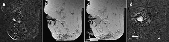
Fig. 69.10 a-d MR-guided vacuum core biopsy.
a Subtraction image showing lesion.
b Coaxial needle.
c Biopsy area after removal of tissue specimens.
d Final subtraction image after further contrast administration showing enhancement due to bleeding in the resection cavity.
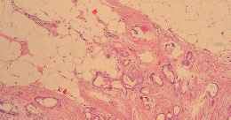
Fig. 69.11 Histology of the left breast: tubular carcinoma.
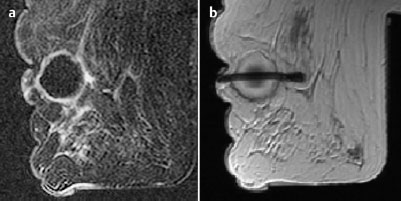
Fig. 69.12 a,b Preoperative hook-wire localization of the lesion between the outer quadrants of the left breast after core biopsy.
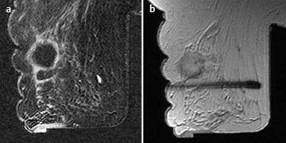
Fig. 69.13 a-c Preoperative hook-wire localization of the second lesion between the inner quadrants of the left breast.
Histology
Tubular carcinoma (diameter 7 mm).
TC pT1 b, pNO, G1 left lateral + fibroadenoma left medial.
Stay updated, free articles. Join our Telegram channel

Full access? Get Clinical Tree


