Midsagittal T2WI.

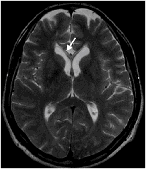
Axial T2WI.
Marchiafava-Bignami Disease
Primary Diagnosis
Marchiafava-Bignami disease
Differential Diagnoses
Partial callosal agenesis
Chronic anterior cerebral artery infarction
Susac syndrome
Multiple sclerosis
Imaging Findings
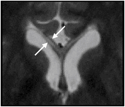
Fig. 69.1: Midsagittal T2WI showed marked thinning of anterior corpus callosum (CC), particularly involving the genu and the body (arrow). The remainder of the CC portions is normal. Fig. 69.2: Axial T2WI demonstrated marked anterior CC thinning with normal-appearing posterior portions. Fig. 69.3: Zoomed in axial T2WI showed that the thinned genu of the CC has a three-layered appearance. The outer layers indicated that the gliotic white matter and the inner portion are as hyperintense as the CSF.
Discussion
The presence of anterior callosal thinning and the three-layered appearance of the CC are typical for Marchiafava-Bignami (MB), particularly if there is a history of chronic alcoholism/alcohol consumption. In cases of partial callosal agenesis, the missing portions of the CC are typically posterior, usually the splenium and/or rostrum. In such situations, the third ventricle is usually high-riding, the lateral ventricles are parallel (non-converging) and colpocephaly, and Probst bundles are evident. In this patient, the diagnosis of partial callosal agenesis is unlikely because the abnormal portions of the CC are anterior, and thinned as opposed to missing. The remaining accompanying findings compatible with partial callosal agenesis are absent, excluding it as the primary diagnosis.
Isolated CC infarcts are rare, possibly because of the rich blood supply from three main arterial systems: the anterior cerebral, anterior communicating, and posterior cerebral arteries. The body of the CC is usually supplied by the pericallosal branch of the anterior cerebral artery. The subcallosal and medial callosal arteries, branches of the anterior communicating artery, usually supply the anterior portion of the CC. The posterior pericallosal artery, a branch of the posterior cerebral artery, usually supplies the splenium of the CC. If cerebral infarct was the causative agent of the imaging findings in this patient, two different arterial territories would have been involved, which makes the diagnosis of CC chronic infarct less likely. Moreover, isolated CC infarct would be an unlikely event in a patient with no cardiovascular risk.
Susac syndrome (SS) (see Part II: Case 26) and multiple sclerosis (MS) are diseases that typically involve the CC and therefore can cause callosal thinning. Susac syndrome virtually always involves the CC and patients have a typical clinical presentation of encephalopathy, bilateral hearing loss, and branch retinal artery occlusions. Susac syndrome callosal lesions are T2 hyperintense and typically involve the body and splenium of the CC, particularly its middle layers. Typical MS patients have a less severe clinical evolution, when compared to SS patients. Imaging findings include multiple perpendicular callososeptal T2 hyperintensities, and Dawson fingers (areas of dysmyelination, glial scars, or sclerosis) can be seen. Additionally, periventricular, subcortical and deep white matter hyperintensities as well as brainstem, cerebellum, and spinal lesions can be seen. In more advanced cases, diffuse CC thinning can be seen. Owing to the lack of clinical and imaging concordant findings, the possibility of SS and MS are less likely in this case.
Marchiafava-Bignami is a rare, progressive neurologic disease first described in Italian red wine drinkers by Carducci in 1898 and by Marchiafava and Bignami in 1903. It is more commonly seen in middle-aged or elderly alcoholic males. Rarely is it associated with various nutritional deficiencies, as opposed to alcoholism or alcohol abuse. It has been reported that MB individuals are typically heavy wine drinkers, with daily consumption of at least two liters per day for more than 20 years.
The blueprint of MB is demyelination and necrosis of the CC with subsequent atrophy. Clinically, MB patients can present during one of three different stages (acute, subacute, or chronic). In the acute stage, patients may present seizures, impaired consciousness and cognition, gait disturbance, hemiparesis, stupor, coma, and death. Subacute stage features include variable mental confusion, dysarthria, behavioral abnormalities, memory deficits, interhemispheric disconnection states, and impaired gait. Chronic MB is less common and presents as mild dementia, which progresses over a period of several years. As patients in the acute stage usually have a fatal outcome (if left untreated), early diagnosis and treatment are crucial.
The CC is usually affected in the middle portion (middle lamina), its highest myelinated component. Extracallosal structures can also be involved, in particular: the anterior and posterior commissures, the central semiovale, and other major white matter tracts. Other less commonly involved structures are the optic chiasm and tracts, putamen, cerebellar peduncles, and rarely, the cortical gray matter with laminar necrosis, and U fibers. Antemortem recognition of MB is mainly a neuroradiology diagnosis, since the clinical picture is often quite variable. It is important to remember that patients with acute MB can rapidly progress to death.
On imaging, one can appreciate the sparing of the external layers of the CC and destruction of its middle component that gives rise to the three-layered sandwich sign, the characteristic imaging feature of MB. Cystic-necrotic changes are usually seen in the genu and splenium of the CC.
On CT, the involved portions of the CC are hypodense. Occasionally, in the presence of hemorrhage in the subacute stage, lesions can appear hyperdense. On MRI, the inner portion of the CC is hypointense on T1 and hyperintense on T2-weighted and on FLAIR images.
In the acute and subacute stages, one can appreciate thickening of the CC due to edema. In those stages, T2-weighted imaging reveals increased signal in the CC, due to edema and myelin damage. When myelin injury persists with subsequent myelin loss, the CC remains hyperintense in T2WI. In some patients, signal changes can recover to a normal appearance, despite total demyelination. In the chronic stage, there may be development of cystic changes that are T1 hypointense and T2 hyperintense. Sagittal FLAIR images are more sensitive to depict these chronic changes. The middle portion of the CC shows hypointense signal, reflecting white matter necrosis and leukomalacia; external layers show hyperintense gliotic rim.
Diffusion-weighted imaging is useful to detect areas of active demyelination, which are seen as areas of restricted diffusion. Positive restricted diffusion during the acute stage does not always indicate permanent, irreversible tissue damage, as sometimes those signals may regress with no apparent permanent damage. Owing to acute stage myelin loss, the expected findings on spectroscopy are an increase in choline peaks and an increase in the choline/creatine ratio. Lactate peaks are expected in the acute/subacute stages of demyelination. Choline and lactate peaks and choline/creatine ratios can return to normal after clinical improvement.
Diffusion tensor imaging is important to demonstrate regional abnormalities in the CC when there are no evident changes demonstrated on conventional MRI. Fiber tracking can demonstrate disruption of the axonal fibers within the CC, particularly in the middle portion of the body. Currently, there is no standard protocol for MB treatment; however, the patients are usually treated with thiamine, folate, and vitamin B complex, with good clinical improvement in many cases. Acute MB can be considered a neuroradiology emergency in which early recognition and treatment are critical for satisfactory outcome.
Stay updated, free articles. Join our Telegram channel

Full access? Get Clinical Tree


