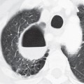
CASE 69 53-year-old woman status post kidney transplant eight months earlier with five-day history of vesicular skin rash followed by progressive dyspnea, cough, tachypnea, and chest pain PA (Fig. 69.1A) and lateral (Fig. 69.1B) chest radiographs show multiple subcentimeter nodules of varying size distributed throughout both lungs. Chest CT (lung window) (Figs. 69.1C, 69.1D, 69.1E, 69.1F) reveals 1–10 mm well-defined and ill-defined nodules randomly disseminated bilaterally. At least one nodule in the left lower lobe has surrounding ground glass (Fig. 69.1E). Note the absence of pleural effusion and lymphadenopathy. Varicella-Zoster Pneumonia • Cytomegalovirus (CMV) • Adenovirus • Measles • Respiratory Syncytial Virus (RSV) • Disseminated Fungal Pulmonary Infection • Mycobacterium tuberculosis • Metastatic Pulmonary Disease Varicella-zoster virus (VZV) is a contagious herpes virus that can cause two distinct clinical syndromes. The first, varicella (chickenpox), is most commonly a self-limited mucocutaneous process that occurs primarily in children, and less often in adults. The second, herpes zoster (shingles
 Clinical Presentation
 Radiologic Findings
 Diagnosis
 Differential Diagnosis
 Discussion
Background
![]()
Stay updated, free articles. Join our Telegram channel

Full access? Get Clinical Tree


