7

Case 7


Indication: Screening.


History: Unremarkable.


Risk profile: Bilateral breast cancer in mother at 45 and 65 years.


Age: 41 years


image


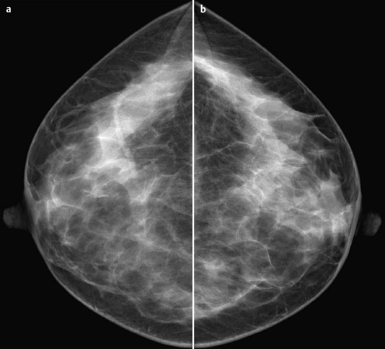
Fig. 7.1 a,b Digital mammography, CC view.


image


Clinical Findings


No pathological findings on inspection or palpation.


Ultrasound


No pathological findings.


image


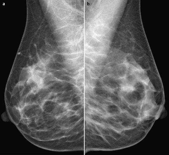
Fig. 7.2a,b Digital mammography, MLO view.


image


image


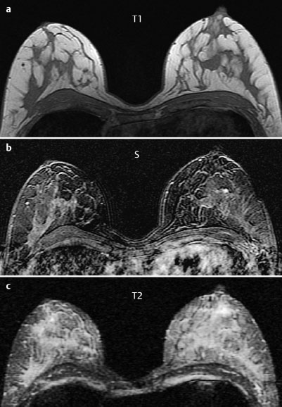
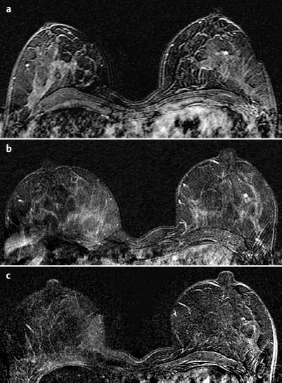
Fig. 7.3a–c Contrast-enhanced MRI of the breasts.


image


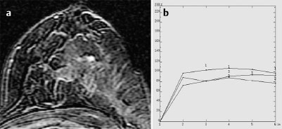
Fig. 7.4a,b Signal-to-time curves.


image


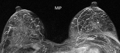
Fig. 7.5 Contrast-enhanced MR mammography. Maximum intensity projection.


image


Please characterize mammography and MRI findings.


What is your preliminary diagnosis?


What are your next steps?


These examinations were performed in a screening situation in a woman with an increased familial risk.


Ultrasound


Ultrasound showed no pathological findings. In particular, there was no circumscribed lesion corresponding to the enhancing area in the left breast seen in MRI.


Mammography


Digital mammograms demonstrated bilaterally symmetric, extremely dense parenchymal structures, ACR type 4. Under these limiting conditions, no lesions or masses were visible. No calcifications (BI-RADS right 1/left 1). PGMI: CC view P; MLO view P.


MR Mammography


MRI depicted a 5 mm mass in the central part of the left breast with increased vascularization. No other findings.


MRI Artifact Category: 3


MRI Density Type: 2


MRM score


Finding


Points


Shape


round


0


Border


well-defined


0


CM Distribution


inhomogeneous


1


Initial Signal Intensity Increase


moderate


1


Post-initial Signal Intensity Character


plateau


1


MRI score (points)


3


MRI BI-RADS


3


image Differential Diagnosis


Fibroadenoma, adenoma, papilloma, small carcinoma.


BI-RADS Categorization


Clinical Findings


right 1


left 1


Ultrasound


right 1


left 1


Mammography


right 1


left 1


MR Mammography


right 1


left 3


BI-RADS Total


right 1


left 3


Procedure


Follow-up examination (MRI) in 6 months.


image


Fig. 7.6a–c Monitoring of findings.


a First examination.


b Second examination 6 months after first MRI.


c Second examination 12 months after first MRI.


Sep 3, 2016 | Posted by in GENERAL RADIOLOGY | Comments Off on 7

Full access? Get Clinical Tree
Get Clinical Tree app for offline access