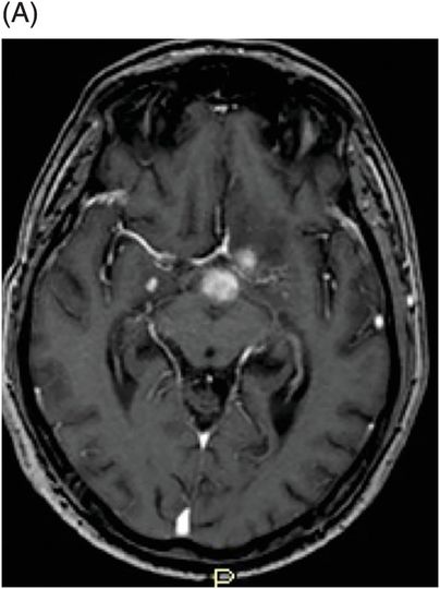
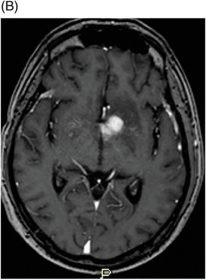
(A) Postcontrast axial T1WI sequence through same level as Fig. 7.1A. (B) Postcontrast axial T1WI through the same level as Fig. 7.1B.
Coronal postcontrast T1WI sequence through the hypothalamus.
Whipple Disease with Central Nervous System Involvement
Primary Diagnosis
Whipple disease with central nervous system involvement
Differential Diagnoses
Paraneoplastic limbic encephalitis
Pilomyxoid astrocytoma
Metastasis
Imaging Findings
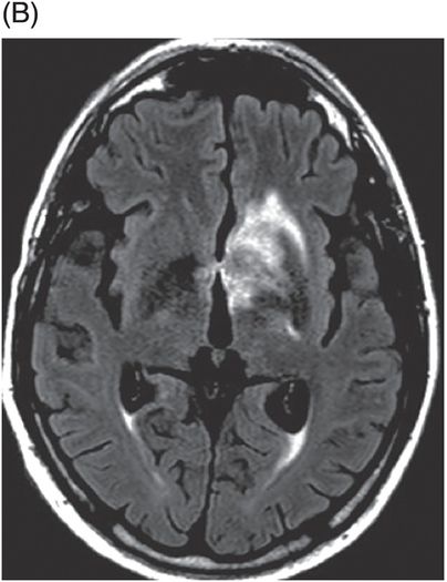
Fig. 7.1: (A) Axial FLAIR image through the level of the cerebral peduncles demonstrated increased FLAIR signal involving the left inferior frontal lobe, bilateral medial temporal lobes, and inferior hypothalamus. Mass effect is absent. (B) Axial FLAIR image at slightly higher level demonstrated increased FLAIR signal involving the left inferior frontal lobe, left hypothalamus, and left inferior basal ganglia. Subtle signal abnormality in the right inferior basal ganglia was also noted. Fig. 7.2: (A) Postcontrast axial T1WI sequence through same level as Fig. 7.1A demonstrated nodular enhancement of the left inferior frontal lobe and the hypothalamus. (B) Postcontrast axial T1WI sequence at the same level as Fig. 7.1B demonstrated nodular enhancement involving the left inferior basal ganglia and the hypothalamus. Fig. 7.3: Coronal postcontrast T1WI sequence through the hypothalamus demonstrated left-predominant nodular enhancement of the hypothalamus. In addition, subtle nodular enhancement was noted in the right medial temporal lobe, right basal ganglia, and in the left cingulum.


Stay updated, free articles. Join our Telegram channel

Full access? Get Clinical Tree


