7 Musculoskeletal
Approach
Most musculoskeletal examinations obtained in the emergency department are requested for characterization of fractures, evaluation of joint effusions, exclusion of osteomyelitis in patients with cellulitis, and detection of foreign bodies in lacerations and puncture wounds. For most osseous injuries, plain radiographs are sufficient for diagnosis, and CT is reserved for more precise characterization of complex fractures (including tibial plateau and calcaneal fractures), localization of intraarticular bone fragments, and detection of soft tissue abscess or gas. MRI is often the best choice for evaluation of cartilaginous, meniscal, and ligamentous injuries, but it is usually not necessary (or even available) in the emergency department.
While one should always evaluate every bone included on a radiograph, it is very helpful to know the exact location of the patient′s pain as well as the mechanism of injury. Fractures can be extremely subtle, and degenerative changes or remote injuries can simulate acute fractures. Accurate clinical information reduces perceptual failures and allows more accurate assessment of a borderline finding.
“Satisfaction of search” is a well-known cause of error among radiologists; after identifying one fracture, it is important to make sure that no other fractures are present. Another common cause of diagnostic error in interpreting bone radiographs is failing to obtain adequate orthogonal and (if necessary) supplemental views, either because the patient was unable to cooperate or incorrectly positioned, or because the optimal study for the clinical problem was not ordered.
Finding an area of focal soft tissue swelling or poor subcutaneous fat definition should increase suspicion for an adjacent subtle fracture. A joint effusion, particularly in the knee or elbow, is another important indirect sign of a fracture.
Long-bone fractures should be described concisely, using consistent descriptive and anatomic terms and addressing fracture location, configuration, and alignment. Although exact classification and most measurements are usually best left to the treating clinician, the radiologic interpretation should provide all descriptive information necessary to categorize a fracture for accurate orthopedic diagnosis and management.
Location
Proximal
Diaphyseal
Metaphyseal
Distal
Intra-articular
Configuration
Compound (open)
Simple (transverse, oblique, or spiral)
Segmental
Comminuted
Avulsed
Impacted
Osteochondral
Torus (buckle)
Incomplete (greenstick)
Alignment
Displacement
Distraction
Override
Angulation
Radiographs are often obtained in patients with cellulitis or skin ulcer in order to exclude osteomyelitis. Most bone radiographs obtained in this setting are normal or show sequelae of underlying diabetes mellitus such as vascular calcification or neuropathic arthropathy. Radiographic findings that indicate bone infection include focal cortal loss, periosteal new bone formation, and, less commonly, sclerosis or an endosteal lytic lesion. MRI and radionuclide bone scan are more sensitive for detection of bone infection but need not be urgently obtained.
In patients with lacerations and puncture wounds, ultrasound or radiographs can localize foreign bodies. Sensitivity for detection depends on the size and density of the object. Metal, glass, stone, and bone fragments are usually radio-opaque and often localizable by radiographs or CT. In contrast, wood splinters and plastic are usually not seen on plain radiographs but, if large enough, can be found using high-frequency ultrasound. Dry wood (as in a pencil) may be visible on CT as a linear body with the approximate attenuation of air.
Imaging
Shoulder
Radiographs
AP internal rotation
AP external rotation
Axillary and/or trans-scapular
Checklist
Scapula body
Acromion
Coracoid process
Clavicle
Humeral head
Glenohumeral, acromioclavicular, and coracoclavicular articulations
Common Injuries
Anterior dislocation
Posterior dislocation
Acromioclavicular separation
Clavicle fracture
Humeral head/neck fractures
Elbow
Radiographs
AP
Lateral
Angled oblique (Greenspan)
Checklist
Humerus
Ulna
Radius
Effusion (anterior and posterior fat pad signs)
Position and appearance of ossification centers (in children)
Radiocapitallar and anterior humeral line alignment (in children)
Common Injuries
Dislocation
Radial head fracture
Coronoid process fracture
Olecranon fracture
Humeral supracondylar fracture
Humeral epicondylar avulsion
Hand and Wrist
Radiographs
AP
Lateral
Oblique
Scaphoid (wrist)
Checklist
Soft tissues
Distal radius and ulna
Carpal bones
Metacarpals
Phalanges
Carpal arch alignment (AP)
Lunate-capitate alignment (lateral)
Common Injuries
Distal radius/ulnar styloid fracture
Scaphoid fracture
Lunate and perilunate dislocation
Triquetral fracture
Metacarpal fracture
Dorsal and volar plate avulsion fractures
Amputations
Pelvis
Radiographs
AP
Judet (bilateral oblique)
Inlet and outlet
Checklist
Pubic rami, pubic symphysis, and ischial tuberosities
Acetabula
Femoral heads and necks
Iliac bones
Sacral alae and sacroiliac articulations
Lower lumbar vertebrae/transverse processes
Pelvic soft tissues
Common Injuries
Lateral compression fracture
Anterior-posterior compression fracture/open-book fracture
Windswept pelvis
Vertical shear fracture
Pubic ramus fracture
Acetabular fracture
Hip
Radiographs
AP
Cross-table lateral
Frog-leg lateral
Checklist
Pubic rami, pubic symphysis, and ischial tuberosities
Acetabula
Femoral heads and necks
Iliac bones
Pelvic soft tissues
Common Injuries
Subcapital, transcervical, and basicervical femoral neck fracture
Intertrochanteric or subtrochanteric fracture
Greater or lesser trochanter avulsion
Dislocation
Pubic ramus fracture
Acetabular fracture
Slipped capital femoral epiphysis (children)
Knee
Radiographs
AP
Cross-table lateral
Oblique
Patellar (sunrise)
Checklist
Soft tissues
Patella
Quadriceps and patellar tendons
Effusion or lipohemarthrosis
Femoral condyles
Tibial plateau and spines
Fibular head
Common Injuries
Patellar fracture
Quadriceps or patellar tendon rupture
Anterior cruciate ligament injury (look for lateral condylar notch sign)
Tibial plateau fracture
Tibial spine avulsion
Segond fracture (associated with ACL tear)
Proximal fibular avulsion fracture (arcuate sign, associated with posterolateral ligamentous injury)
Ankle and Foot
Radiographs
AP
Lateral
Oblique
Calcaneus
Checklist
Soft tissues
Distal fibula
Medial tibial malleolus
Cortex of distal tibia and talar dome (look for osteochondral fractures)
Posterior tibia (lateral view)
Tibiotalar interval and ankle mortise congruity
Talus
Calcaneus and midfoot (navicular, cuboid, cuneiforms)
Base of fifth metatarsal
Alignment of metatarsals with respect to cuneiforms
Metatarsal shafts (stress fractures)
Phalanges
Common Injuries
Rotational ankle injuries (distal fibular, bimalleolar, and trimalleolar fractures)
Axial load (pilon) fractures of the distal tibia
Salter Harris fractures in children and adolescents (triplane fracture, Tillaux fracture)
Calcaneal fractures
Lisfranc fracture dislocations
Metatarsal stress fractures
Fifth metatarsal fractures (Jones, Pseudo-Jones)
Extremity Computed Tomography
Indications: Complex fractures involving the elbow, tibial plateau, ankle, and calcaneus. Exclusion of intra-articular fracture fragments.
Technique: 150 mA, 120 kV
Images: 2.5-mm axial with 0.6-mm reconstruction and 2-mm coronal and sagittal reformations, optional 3D reformations
Lower Extremity CT Angiography
Indications: Suspected lower extremity vascular injury.
Technique: 150 mA, 120 kV
IV contrast: 1.5 mL/kg at 5 mL/sec with 30 mL saline chaser. Timing bolus (20 mL) plus 5 sec or empiric 25-second delay
Images: 2.5-mm axial with 0.6-mm reconstruction and 2-mm coronal and sagittal reformations, optional 3D reformations. Image both lower extremities.
Upper Extremity CT Angiography
Indications: Suspected upper extremity vascular injury.
Technique: 150 mA, 120 kV
IV contrast: 1.5 mL/kg at 5 mL/sec with 30 mL saline chaser. Timing bolus (20 mL) plus 5–8 second delay
Images: 2.5-mm axial with 0.6-mm reconstruction and 2-mm coronal and sagittal reformations, optional 3D reformations. The arm may be positioned on the side, which results in less motion but more noise, or above the head, which has less noise but more motion.
Orthopedic Hardware
While the variety of orthopedic screws, plates, prostheses, and other appliances is broad and may be daunting, it is helpful to be able to describe the more commonly seen hardware ( Fig. 7.1 ).


Clinical Presentation and Differential Diagnosis
Clinical Presentations and Appropriate Initial Studies
Soft Tissue Swelling
Radiograph
Ultrasound
CT for detection of subtle air if necrotizing fasciitis is considered
– Cellulitis
– Soft tissue abscess
– Necrotizing fasciitis
– Deep venous thrombosis
– Foreign body
Atraumatic Pain
Radiograph
– Primary bone malignancy
– Metastasis
– Osteomyelitis
– Osteoid osteoma
– Stress fracture
– Hardware failure or loosening
Arthropathy
Radiograph
– Inflammatory arthritis
– Septic arthritis
– Degenerative arthropathy
– Gout or other crystal-induced disease
– Posttraumatic arthropathy
– Neuropathic arthropathy
Differential Diagnosis
Multiple Lytic Lesions
Metastases
Multiple myeloma
Lymphoma
Destructive Bone Lesion
Metastasis
Primary bone tumor
Lymphoma
Plasmacytoma
Eosinophilic granuloma
Osteomyelitis
Intramedullary Lesion with Chondroid Matrix
Enchondroma
Bone infarct
Chondrosarcoma
Sclerotic bone lesion
Osteoblastic metastasis (breast, prostate)
Bone island
Paget disease
Lymphoma
Benign-Appearing Expansile Lesion
Fibrous dysplasia
Solitary bone cyst
Aneurysmal bone cyst
Giant cell tumor
Monoarthropathy
Osteoarthritis
Gout
Rheumatoid arthritis
Septic arthritis
Tuberculosis
Trauma
Arthritis with Osteopenia
Rheumatoid arthritis
Juvenile rheumatoid arthritis
Lupus
Septic arthritis
Tuberculosis
Arthritis with Normal Bone Density
Osteoarthritis
Gout
Calcium pyrophosphate deposition (CPPD)
Psoriatic
Ankylosing spondylitis
Neuropathic arthropathy
Destructive Arthropathy
Rheumatoid arthritis
Juvenile rheumatoid arthritis
Psoriatic arthritis
Neuropathic arthropathy
Sacroiliitis
Ankylosing spondylitis
Inflammatory bowel disease
Psoriatic arthritis
Reiter syndrome (asymmetric)
Arthritis—Hands and Feet
Osteoarthritis
Erosive osteoarthritis
Rheumatoid arthritis
Psoriatic arthritis
Gout (foot)
Arthritis—Shoulders and Hips
Rheumatoid
Crystal arthropathy
Collagen vascular disease
Scapular Fracture
Scapular fractures result from direct impact to the shoulder and are usually associated with high-energy mechanisms. Patients with a scapular fracture are consequently likely to have associated torso injuries including pneumothorax, pulmonary contusion, rib or vertebral compression fractures, upper and lower extremity fractures, and upper extremity neurovascular structures (axillary artery and nerve, brachial plexus).
Scapular fractures may be difficult to diagnose on conventional radiographs but are easily appreciated on chest CT obtained in polytrauma. They are described as body, spine, acromion, coracoid, scapular neck, and glenoid fractures. Fragment displacement is usually minimal due to the supporting muscles and periosteum. Unless the glenoid fossa is involved, most scapular fractures are managed nonoperatively with a sling and outpatient orthopedic evaluation. Fractures that extend to the articular surface may require operative reconstruction ( Fig. 7.2 ).

Acromioclavicular Separation
Acromioclavicular separation can result from a direct blow to the shoulder or a fall onto the shoulder with the arm adducted. These mechanisms force the scapula inferiorly and medially with respect to the distal clavicle. In the case of a fall on the outstretched hand, the scapula is transiently displaced superiorly from the clavicle, injuring the acromioclavicular ligament.
The inferior cortex of the acromion and distal clavicle should normally align on the AP view. The distance between the acromion and distal clavicle is variable but usually less than 8–10 mm. Weight-bearing views with comparison to the uninjured shoulder may be necessary to demonstrate subluxation.
Grade I: Normal or slight acromioclavicular subluxation. Acromioclavicular ligament sprain with intact coracoclavicular ligament.
Grade II: Acromioclavicular ligament tear with acromioclavicular widening or distal clavicular elevation. Coracoclavicular ligament injury without widening of the normal coracoclavicular distance (< 1.3 cm).
Grade III: Disruption of both acromioclavicular and coracoclavicular ligaments. Acromioclavicular subluxation with elevation of distal clavicle relative to the acromion. Coracoclavicular distance > 1.3 cm or a side-to-side difference of > 5 mm on bilateral AP views. Weight-bearing radiographs may be necessary to reveal these findings.
Grade I and II injuries are usually treated conservatively. Grade III injuries may benefit from operative stabilization ( Fig. 7.3 ).

Anterior Shoulder Dislocation and Luxatio Erecta
Anterior shoulder dislocation is the most common type of shoulder dislocation (~ 95%) and occurs with forced arm abduction, external rotation, and extension. The humeral head is displaced anterior, medial, and inferior to its normal location, and its posterolateral surface strikes the antero-inferior surface of the scapular glenoid. In anterior dislocation, impaction fractures of the posterolateral humeral head (Hill-Sachs lesion) and anteroinferior glenoid labrum avulsion (Bankart lesion) commonly occur. An osseous glenoid rim fracture, when present, is referred to as a bony Bankart lesion.
Pain and muscle spasm are the rule, and patients typically hold the affected arm in slight abduction and external rotation.
Anterior shoulder dislocations are well characterized using a standard trauma series consisting of AP views in internal and external rotation, the scapulary view, and the axillary view. CT or MR may be useful for evaluation of osteocartilaginous fractures or intra-articular fragments. MR, in particular, is superior for identification of rotator cuff, capsular, and glenoid labral injuries.
Luxatio erecta (inferior dislocation) is an uncommon variant of anterior dislocation and tends to occur in elderly individuals. The mechanism of injury is forceful hyperabduction with impingement of the humeral neck on the acromion. In this injury the arm is held upward or behind the head. The displaced humeral head is often palpable on the lateral chest wall.
The AP radiograph is diagnostic; the humeral head is inferiorly displaced with the humeral shaft directed superolaterally along the glenoid margin. The humeral articular surface is directed inferiorly and is no longer in contact with the inferior glenoid rim.
Luxatio erecta is almost always accompanied by detachment of the rotator cuff and neurovascular compression. Associated fractures are also common but difficult to detect clinically because of severe shoulder pain. Early reduction should be attempted in an effort to prevent any neurologic or vascular damage. In most cases, reduction is not difficult and may be accomplished by the use of traction-counter-traction maneuvers ( Fig. 7.4 ).

Posterior Shoulder Dislocation
Posterior shoulder dislocation is much less common than anterior shoulder dislocation (~ 5%) and can be a difficult diagnosis both clinically and radiographically. Posterior dislocations follow seizures, electrocution, or a blow to the back of the shoulder with the arm internally rotated and abducted. Patients are usually unable to rotate their arm externally but do not present with striking deformity, and these injuries can be missed if axillary or scapulary views are not obtained.
On frontal radiographs, the normal superposition of humeral head on the glenoid and the humeral profile in external rotation, which demonstrates the greater tuberosity, is lost. Because the humeral head is held in internal rotation, it appears rounded (the “lightbulb” sign). Most patients cannot externally rotate the affected arm, and standard internally and externally rotated AP radiographs appear identical. A fracture of the anterior humeral head, known as the trough line or reverse Hill-Sachs lesion, reflects impaction of the anterior humeral head against the posterior glenoid rim. The corresponding fracture of the posterior glenoid (when present) is called a reverse Bankart lesion.
Reducing a posterior shoulder dislocation is generally more difficult than reducing an anterior dislocation; if available, orthopedic consultation is indicated prior to reduction, as is adequate sedation, analgesia, and muscle relaxation ( Fig. 7.5 ).

Humeral Head Fracture
Humeral head and neck fractures are typically seen in elderly women after a fall on an outstretched hand. In younger patients, this injury is a consequence of high-energy trauma and is usually associated with other significant injuries. Patients present with shoulder pain, swelling, and tenderness; crepitus; and ecchymosis. Sensory disturbance, paresthesia, and diminished pulses indicate associated axillary nerve or artery injury.
Radiographs should be obtained in AP, transscapular, and (if possible) axillary views. Articular surface fractures may be associated with a hemarthrosis that displaces the humeral head inferiorly (pseudosubluxation).
The Neer classification system divides the proximal humerus into four parts, which are located between the epiphyseal lines where fractures primarily occur: the anatomic neck, the surgical neck, and the greater and lesser tuberosities. Fragments are considered displaced if separated by more than 1 cm or > 45° angulation. A one-part fracture contains no displaced fragments, regardless of the number of fracture lines. Two-part, three-part, and more severely comminuted fractures are characterized by progressively greater displacement and angulation of the small fragments.
One-part fractures are treated with immobilization and analgesics, but all other proximal humeral fractures require urgent orthopedic consultation in the emergency department because of the high risk of complications. Closed reduction, operative fixation, or a combination of the two may be necessary ( Fig. 7.6 ).

Elbow Dislocation
Elbow dislocations are common, second in frequency only to shoulder and finger dislocations. Ninety percent are posterior and due to a fall on an extended, abducted arm. Clinically, the elbow is flexed at 45° with associated swelling, and the posteriorly dislocated olecranon process is easily palpated in its abnormal position.
Simple dislocations are treated with closed reduction and brief immobilization. Postreduction images should be carefully evaluated for radial head fracture, coronoid process fracture, or intra-articular fragments; CT may be helpful for complex or comminuted injuries. Complex fracture-dislocations and unstable dislocations require operative treatment.
The “terrible triad” refers to elbow dislocation, usually under varus stress, with associated fractures of the ulnar coronoid process and radial head. In this injury, the lateral collateral ligament is almost always disrupted, resulting in an unstable elbow. It is generally managed surgically by reattaching the ulnar coronoid process and affixing the radial head fracture (or replacing the radial head).
Complex fracture/dislocations of the elbow are more likely to be complicated by osteoarthritis, range of motion limitation, instability, and recurrent dislocation ( Fig. 7.7 ).

Radial Head Fracture and Essex-Lopresti Fracture-Dislocation
Radial head fractures, the most common of the adult elbow fractures, are due to falls on an outstretched hand in which the radial head is driven into the humeral capitellum. Associated injuries are common and include coronoid fracture, elbow dislocation, medial collateral ligament injury, interosseous membrane injury, and damage to the triangular fibrocartilage complex at the wrist.
Passive pronation and supination of the forearm are painful, especially over the lateral elbow. Most fractures are subtle and reflect impaction at the radial neck. In the setting of acute trauma, a joint effusion, even if there is no cortical disruption or contour deformity, indicates a nondisplaced radial head fracture and should be treated accordingly. Signs of an effusion include elevation of the anterior elbow fat pad (sail sign) and visibility of the posterior olecranon fat pad.
The Mason classification is summarized in Table 7.1 .
Type I (nondisplaced) fractures are treated conservatively with brief immobilization and analgesia. Displaced or otherwise complicated fractures, and those with restricted range of movement, should be referred to an orthopedic surgeon. Type II injuries are treated with open reduction and internal fixation. Type III injuries often require excision of the radial head and prosthetic replacement.
The Essex-Lopresti fracture-dislocation is defined by interosseous ligament disruption, usually accompanied by a fracture of the radial head and disruption of the triangular fibrocartilage of the wrist. The result is a dislocation of the distal radioulnar joint, causing pain in the wrist and forearm, with pronation and supination, and swelling and tenderness over the fractured radial head.
On standard elbow radiographs, radial head fractures may be subtle, and the Essex-Lopresti fracture-dislocation is especially difficult to diagnose. The radio-capitellar line should transect both the radial head and the capitellum on lateral view. Lateral projection is also best for assessment of any distal radioulnar dislocation.
Prompt consultation with orthopedic surgery should be arranged for assessment of both the elbow and distal radioulnar joint. Treatment consists of repair or replacement of the radial head ( Fig. 7.8 ).

Ulnar Olecranon Fracture
Olecranon fractures are common in the elderly, but they can occur at any age. Distal humeral fractures and radial head fractures may be associated with and result in the grossly unstable “floating elbow.”
Best seen on lateral view, the fracture appears as a defect extending from the dorsal olecranon cortex to the articular surface of the trochlea. In most cases, the fracture fragments are widely distracted by the unopposed action of the triceps muscle, which inserts on the olecranon ( Table 7.2 ).
In most cases, olecranon fractures require open reduction and internal fixation to restore the articular surface and preserve the elbow extensor mechanism. Nondisplaced fractures can be managed conservatively ( Fig. 7.9 ).

Forearm Fractures
The Monteggia fracture (or fracture-dislocation) is a proximal ulnar fracture accompanied by radial head dislocation. It can be caused by a direct blow or a fall on an outstretched hand and results in elbow deformity, swelling, and pain with supination or pronation. Radial head dislocation may be subtle; in the normal elbow, a line drawn through the center of the radial head and shaft should intersect the capitellum on all views. Suspect dislocation if this is not the case.
Most pediatric fractures can be managed by closed reduction. Adult fractures usually require operative fixation.
The Galeazzi fracture is a distal radial diaphysis fracture with dislocation of the distal radioulnar joint (DRUJ). Gale-azzi fractures are three times as common as Monteggia fractures. The mnemonic MUGR, which stands for “Monteggia—ulna, Galeazzi—radius,” aids in recalling which bone is fractured in these injuries.
Galeazzi fractures are usually seen in children between 9 and 12 years of age and result from impact to the dorsolateral wrist or a fall onto an outstretched hand.
Radiographic findings include a transverse or oblique distal radial diaphysis fracture, widened DRUJ, and distal subluxation of the ulna relative to the radius.
Urgent operative fixation is normally required for adults with Galeazzi fractures in order to stabilize the fracture and DRUJ. Children younger than 10 years may be treated with closed reduction and splinting, but they should have prompt follow-up orthopedic evaluation.
A nightstick fracture is an isolated mid-shaft ulnar fracture that results from direct trauma to the ulna along its subcutaneous border. In cases of assault, it is a defensive fracture that occurs when a victim attempts to protect his face from an overhead blow. In contrast to the similar-appearing Monteggia fracture, the radiocapitellar relationship is normal.
Nondisplaced fractures are treated with splint immobilization. Open reduction and internal fixation is usually necessary when displacement is greater than 50% or angulation is greater than 10° in any plane ( Fig. 7.10 ).

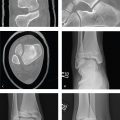
Distal Radius Fractures
Hutchinson fracture is an oblique fracture of the radial styloid that extends into the radiocarpal joint. It is also sometimes called a chauffeur′s fracture, recalling the historical mechanism of injury: a kickback of an early model automobile′s crank handle striking the chauffeur′s wrist.
The fracture is due to axial compression of the scaphoid into the distal radius with radial styloid fracture and radial collateral ligament avulsion. Hutchinson fractures are often associated with scapholunate injury and perilunate dislocation. It is an unstable fracture and requires operative fixation and immobilization.
Colles fracture is an intra- or extra- articular transverse fracture of the distal radius with dorsal displacement and dorsal angulation (apex volar) of the distal fragment, and it is usually due to a fall onto a hyperextended wrist. Clinically, the hand appears to have a “dinner fork” deformity. Colles fractures are more common in elderly women, reflecting demineralization. Most can be managed with closed reduction and cast immobilization with the wrist held in neural to slight flexion.
In contrast to the Colles fracture, the Smith fracture is characterized by volar angulation of the distal fragment. These extra-articular injuries are sometimes called a reverse Colles fracture, reverse Barton, or Goyrand fracture. Smith fractures result from either a direct blow to the back of the wrist or a fall onto a flexed wrist with the forearm in supination. The hand is palmarly displaced with respect to the forearm, resulting in a “garden spade” deformity on physical examination. Less common than Colles fractures, they are usually seen in younger patients with high-energy trauma. Because of its location, the median nerve is susceptible to injury and should be evaluated before and after closed reduction. Smith fractures are unstable and often require open reduction and internal fixation, particularly if there is intra-articular involvement or if the wrist remains grossly unstable after attempted closed reduction.
Plain radiographs (AP, lateral, oblique) of the wrist are sufficient for diagnosis. Distal radius fractures are classified according to (1) extension into the radiocarpal joint, (2) extension to the distal radioulnar articulation, and (3) the presence of an associated ulnar styloid fracture. These features should be included in a description to facilitate the orthopedic surgeon′s ability to make optimal management decisions ( Fig. 7.11 ).

Scaphoid Fracture and Scapholunate Dissociation
Scaphoid fracture is the most common of the carpal fractures and is usually due to a fall on an outstretched hand. Patients present with wrist pain with tenderness over the “anatomic snuffbox,” or dorso-radial aspect of the wrist near the base of the thumb.
The standard wrist radiographic series (PA, lateral, and oblique) may not be diagnostic. The scaphoid view, a frontal radiograph with the wrist in ulnar deviation, elongates the scaphoid and should be obtained if scaphoid injury is suspected. While radiographs with scaphoid view are the best initial examination and will detect most acute fractures, CT and MRI are also highly sensitive studies and can detect subtle fractures and bone contusions in the patient with a negative wrist radiograph. Immobilization with radiographs obtained 1 to 2 weeks after the injury will also sometimes reveal a previously occult fracture.
Scaphoid fractures can be located at the distal pole (10%), waist (70%), or proximal pole (20%). The primary vascular supply to the scaphoid is via the radial artery, two branches of which supply the distal pole and waist of the scaphoid. With no direct arterial supply, the proximal pole depends on fracture union for revascularization. Delayed diagnosis or nonunited fracture may lead to proximal scaphoid avascular necrosis, which appears as sclerosis, fragmentation, and collapse.
Primary treatment is nonoperative with wrist immobilization in slight flexion and radial deviation. Time to union can vary based on location of the fracture and may be as long as 24 weeks for proximal scaphoid fractures. Surgical intervention is reserved for nonunion, displaced, or unstable fractures.
Scapholunate dissociation is the most common ligamentous disruption of the carpus and is often associated with scaphoid fracture. In trauma, the scapholunate ligament fails under high-energy loading of the extended, ulnar-deviated wrist. Scapholunate ligamentous disruption can also be related to chronic arthritis.
On PA radiographs, the normal scapholunate interval should be less than 2 mm. In scapholunate dislocation it is greater than 3 mm.
Pain is localized over the dorsal scapholunate region and exacerbated by dorsiflexion. Scapholunate ligament reconstruction may be required to prevent persistent instability ( Fig. 7.12 ).

Related posts:
Stay updated, free articles. Join our Telegram channel

Full access? Get Clinical Tree


