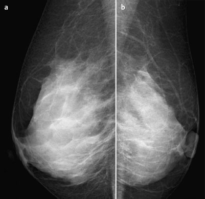
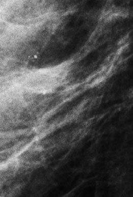
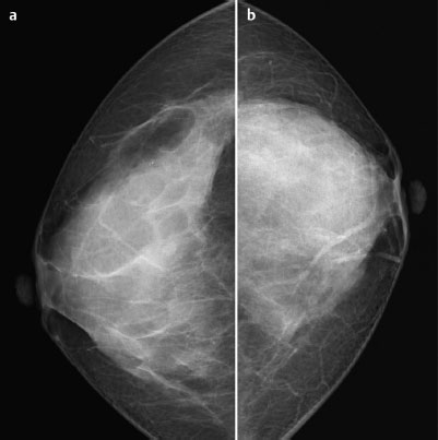
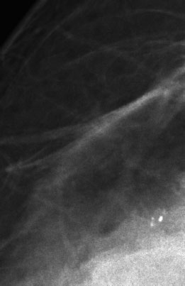
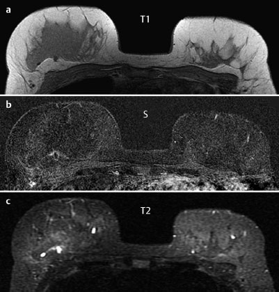
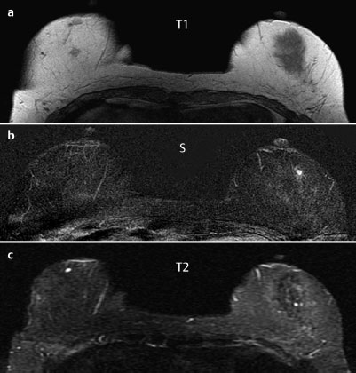
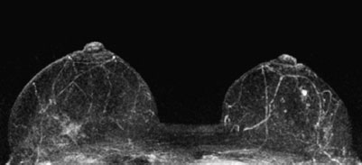
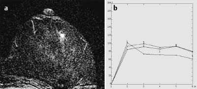
Case 70 Indication: Open biopsy of microcalcifications in the right breast was recommended elsewhere. History: Unremarkable. Risk profile: No increased risk. Age: 49 years. No findings. Normal. Fig. 70.1 a,b Digital mammography, MLO view [imaging not performed by authors]. Fig. 70.2 Digital mammography (MLO), magnification view of the right breast. Fig. 70.3 a,b Digital mammography, CC view [imaging not performed by authors]. Fig. 70.4 Digital mammography (CC), magnification view of the right breast. Fig. 70.5 a-c Contrast-enhanced MRI of the breasts. Fig. 70.6 a-c Contrast-enhanced MRI of the breasts. Fig. 70.7 Contrast-enhanced MR mammography. Maximum intensity projection. Fig. 70.8 a,b Signal-to-time curves. Please characterize the mammography and MRI findings. What is your preliminary diagnosis? What are your next steps? No unusual findings bilaterally. US BI-RADS 1. Mammograms showed bilaterally symmetric extremely dense parenchyma, ACR type 4. In the lower outer quadrant of the right breast, a group of monomorphous microcalcifications was seen. There were no suspicious masses or densities and no architectural distortions. BI-RADS right 3/left 1. PGMI: CC view I (nipple projection); MLO view M (pectoralis muscle, inframammary fold, nipple not in profile). MRI documented a round, ill-defined lesion measuring 6 mm in the left breast behind the nipple, presenting with a ring enhancement, a strong initial signal increase, and postinitial plateau as well as reduced signal in T2-weighted imaging. * Note: The MRI was performed following stereotactic vacuum biopsy on the right breast for the purpose of preoperative local staging. The biopsy region is visible in MRI, in the area of the parenchyma toward the chest wall, as a slight ring enhancement with increased signal in T2-weighted imaging. MRI Artifact Category: 1 MRI Density Type: 1
Clinical Findings
Ultrasound (not shown)

Ultrasound
Mammography
MR Mammography*
MRM score | Finding left | Points |
Shape | round | 0 |
Border | ill-defined | 1 |
CM Distribution | ring | 2 |
Initial Signal Intensity Increase | strong | 2 |
Post-initial Signal Intensity Character | wash-out | 2 |
MRI score (points) |
| 7 |
MRI BI-RADS |
| 5 |
 Differential Diagnosis
Right: DCIS, invasive carcinoma, adenosis, regressive fibroadenoma.
Left: Carcinoma, complicated cyst, fibroadenoma, papilloma, focal adenosis.
BI-RADS Categorization | ||
Clinical Findings | right 1 | left 1 |
Ultrasound | right 1 | left 1 |
Mammography | right 3 | left 1 |
MR Mammography | right 3 | left 5 |
BI-RADS Total | right 3 | left 5 |
Procedure
Stereotactic vacuum biopsy of the calcifications in the right breast (Figs. 70.9 and 70.10).
Histopathology of the right breast
Low grade DCIS.
Further procedure
Bilateral simultaneous MR-guided localization of the region to be excised in the right breast (Fig. 70.11) and the suspicious lesion in the left breast (Fig. 70.12).
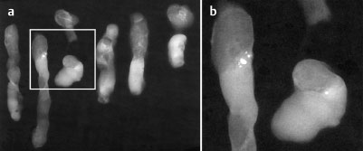
Fig. 70.9 a,b Specimen radiography.
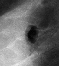
Fig. 70.10 Air-filled cavity at site of biopsy showing complete removal of calcifications.
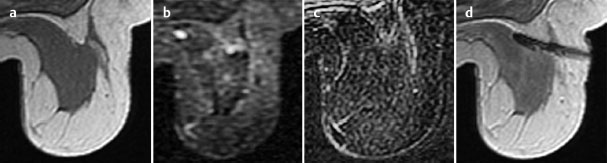
Fig. 70.11 a-d MR-guided localization of the biopsy region in the right breast.
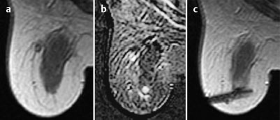
Fig. 70.12 a-c MR-guided localization, left breast.
Diagnosis
Right: DCIS (low grade).
Left: Adenosis.
Stay updated, free articles. Join our Telegram channel

Full access? Get Clinical Tree


