VI Adrenal Glands
CASE 70
Clinical Presentation
A 52-year-old woman complaining of nonspecific abdominal pain.
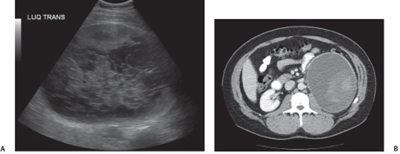
Fig. 70.1 (A) Ultrasound image shows the complex left suprarenal lesion containing multiple septations and mixed hypo-and hyperechoic debris. (B) Axial contrast-enhanced CT image in the same patient shows a large well-defined, thick-walled, complex cystic lesion with hyperdense heterogeneous contents.
Radiologic Findings
Axial computed tomography (CT) and ultrasound images of a left adrenal mass show a large well-defined, thick-walled, complex cystic lesion with heterogeneous contents (Fig. 70.1).
Diagnosis
Left adrenal cyst
Differential Diagnosis
- Adenoma
- Parasitic (hydatid) cyst
- Cystic pheochromocytoma
- Cystic adrenal carcinoma
- Pseudocyst of pancreas, exophytic renal cyst, cystic schwannoma, or cystic adenomatoid tumor mimicking an adrenal cystic lesion
Discussion
Background
Adrenal cystic lesions are more commonly seen in women. It is important to distinguish adrenal cysts from adenoma because adenoma is treated conservatively, whereas adrenal cysts may need surgical intervention if imaging appearances suggest the possibility of complications.
Clinical Findings
Related posts:


Stay updated, free articles. Join our Telegram channel

Full access? Get Clinical Tree


