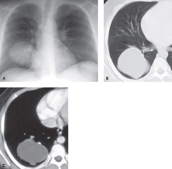
CASE 71 Asymptomatic 35-year-old man PA chest radiograph (Fig. 71.1A) demonstrates a well-marginated polylobular mass in the right lower lobe. Coned-down contrast-enhanced chest CT (lung and mediastinal windows) (Figs. 71.1B, 71.1C) demonstrates a polylobular right lower lobe mass with well-defined borders and intrinsic homogeneous fluid attenuation contents. Note linear peripheral enhancement of the wall of the cystic lesion (Fig. 71.1C). Pulmonary Cystic Hydatid Disease (Echinococcosis) Fig. 71.1 • Pulmonary Bronchogenic Cyst • Primary or Solitary Secondary Malignant Neoplasm The misuse of the term echinococcosis to denote human infection is prevalent, although, by strict definition, echinococcosis refers to infection of non-human carnivores by the adult parasite, while hydatidosis refers to human infection by metacestodes. Pulmonary hydatidosis is a parasitic infectious disease endemic to many parts of the world, particularly underdeveloped sheep- and cattle-raising areas of South America, the Mediterranean region, the Middle East, Africa, and Australia. It is estimated that 65 million individuals are infected worldwide. The life cycle of these organisms requires two hosts: the definitive host, which is a carnivorous dog (or other member of the Canidae or Felidae family) infected with adult egg-producing intestinal tape-worms, and the intermediate host,
 Clinical Presentation
 Radiologic Findings
 Diagnosis

 Differential Diagnosis
 Discussion
Background
![]()
Stay updated, free articles. Join our Telegram channel

Full access? Get Clinical Tree


