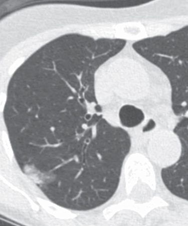
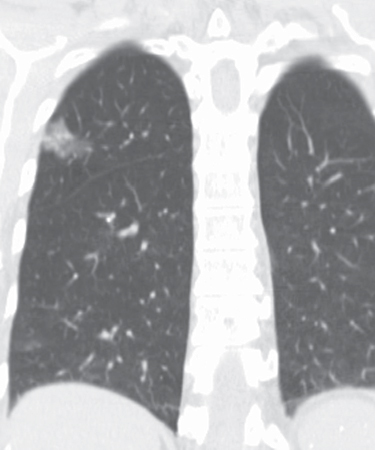
CASE 74 Asymptomatic 77-year-old woman Coned-down unenhanced axial and coronal chest CT (lung window) (Figs. 74.1, 74.2) demonstrates a right upper lobe peripheral subpleural part-solid pulmonary nodule that represents a minimally invasive adenocarcinoma. Coned-down unenhanced chest CT (lung window) of the left lung (Fig. 74.3) shows a 5 mm ground glass left upper lobe nodule presumed to represent a focus of atypical adenomatous hyperplasia based on imaging stability. Lung Cancer; Minimally Invasive Adenocarcinoma • Pulmonary Infection • Adenocarcinoma In Situ; Invasive Adenocarcinoma • Lymphoma Fig. 74.1 Fig. 74.2 The new histologic classification of adenocarcinoma does not include the former bronchioloalveolar carcinoma subtype, which was previously applied to a broad spectrum of neoplasms with widely varied mortality rates and prognostic characteristics. Instead, the new classification introduces the subtypes adenocarcinoma in situ (AIS) and minimally invasive adenocarcinoma (MIA). AIS and atypical adenomatous hyperplasia (AAH) are recognized as precursor or preinvasive lesions for invasive adenocarcinoma of the lung. AIS refers to small lesions (≤3 cm) with pure lepidic growth and no stromal, vascular, or pleural invasion. MIA refers to small lesions (≤3 cm) that exhibit a predominantly lepidic growth and ≤5 mm of locally invasive tumor in any one focus. The term lepidic means “related to scales” or “scaly covering layer” and is used to describe tumors that grow with a replacement pattern or along pre-existing alveolar structures.
 Clinical Presentation
 Radiologic Findings
 Diagnosis
 Differential Diagnosis
 Discussion
Background
Etiology
Related posts:


Stay updated, free articles. Join our Telegram channel

Full access? Get Clinical Tree


