BI-RADS Categorization | ||
Clinical Findings | right 1 | left 1 |
Ultrasound | right 1 | left 3 |
Mammography | right 1 | left 3 |
BI-RADS Total | right 1 | left 3 |
This case demonstrates screening imaging studies of an asymptomatic woman.
Ultrasound
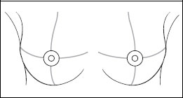
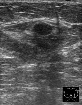
Between the lower quadrants of the left breast there was a round, well-defined lesion with hypoechoic, homogeneous internal texture and unremarkable distal echo pattern. US BI-RADS left (lower quadrants) 3.
Mammography
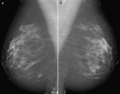
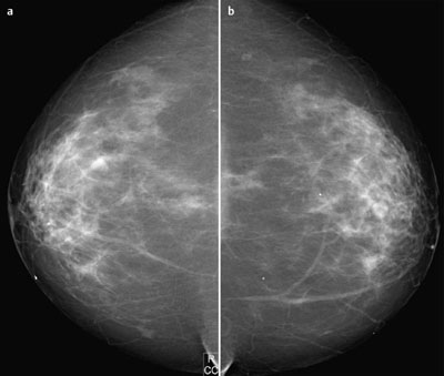
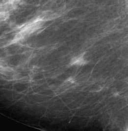
Mammograms showed focally asymmetric, inhomogeneously dense parenchyma, ACR type 3. Between the lower quadrants of the left breast, consistent with the finding in ultrasound, there was a round, well-defined, isodense lesion of 1 cm diameter. In the lower quadrants of the right breast there was a circumscribed density, which in spot compression changed shape and appeared equivalent to glandular tissue. There were no architectural distortions and no suspicious microcalcifications BI-RADS right 3/left 3; after spot compression right 1/left 3. PGMI: CC view P; MLO view P.
 Differential Diagnosis
Left breast: Fibroadenoma, adenoma, papilloma (DD: phyllodes tumor).
Right breast lower quadrants: Isolated parenchymal tissue.
Procedure
A follow-up ultrasound examination of the lesion between the lower quadrants of the left breast was recommended. Screening examinations at intervals of one year should continue.
Diagnosis (without histological confirmation)
Left breast: Fibroadenoma.
Stay updated, free articles. Join our Telegram channel

Full access? Get Clinical Tree


