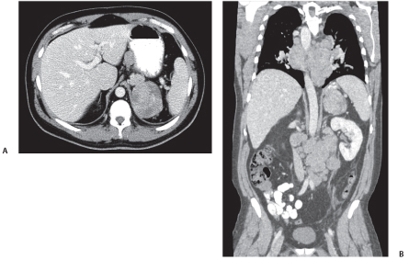
CASE 75 A 40-year-old man presents with a recent-onset cough. Fig. 75.1 (A) Contrast-enhanced axial CT image shows a well-defined rounded, lobulated, heterogeneously enhancing left adrenal mass. (B) Coronal reformatted CT image shows multiple enlarged hypodense celiac and mediastinal lymph nodes with a large heterogeneously enhancing left adrenal mass. Computed tomography (CT) scanning reveals a heterogeneously enhancing rounded left adrenal mass with extensive mediastinal and celiac lymphadenopathy (Fig. 75.1). Adrenocortical carcinoma Adrenocortical carcinoma is a rare malignancy of the adrenocortical tissue, with an incidence of 1 to 2 per million population. It is mostly found in adults, with the median age at diagnosis being 44 years. In 70% of diagnosed cases, tumor invades into adjacent tissues by the time of diagnosis.
Clinical Presentation
Radiologic Findings
Diagnosis
Differential Diagnosis
Discussion
Background
Clinical Findings
Related posts:


Stay updated, free articles. Join our Telegram channel

Full access? Get Clinical Tree


