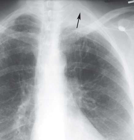
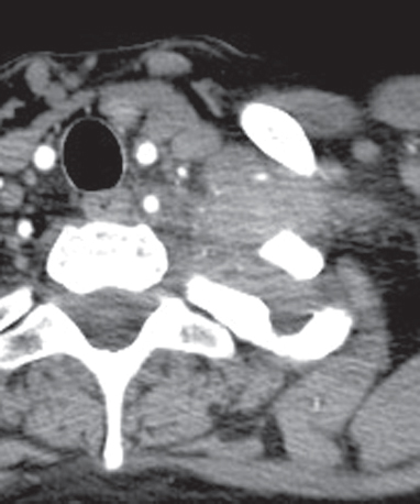
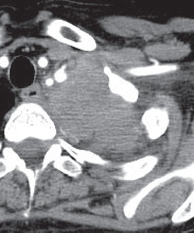
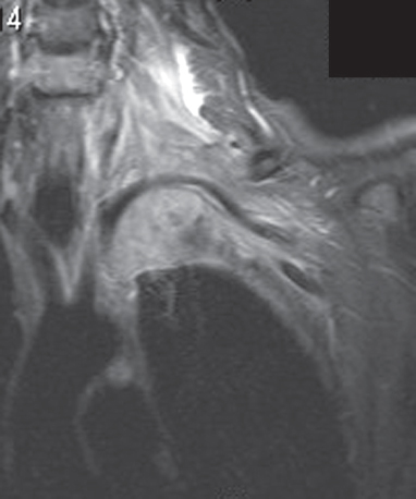
CASE 75 56-year-old woman evaluated because of left upper extremity pain Coned-down PA chest radiograph (Fig. 75.1) shows a left apical lung mass with spiculated borders, associated pleural thickening, and destruction of the left posterior second rib (arrow). Contrast-enhanced chest CT (mediastinal window) (Figs. 75.2, 75.3) demonstrates the locally invasive left apical soft-tissue mass which partially encases the left subclavian artery. Contrast-enhanced coronal T1-weighted MR with fat saturation (Fig. 75.4) reveals to better advantage the extent of the soft-tissue mass, which completely encases the left subclavian artery and surrounds portions of the left brachial plexus Pancoast Tumor; Adenocarcinoma • Pancoast Tumor; Other Cell Type • Pulmonary Lymphoma Fig. 75.1 Fig. 75.2 Fig. 75.3 Fig. 75.4 (Images courtesy of Santiago Martínez-Jiménez, MD, Duke University Medical Center, Durham, North Carolina.)
 Clinical Presentation
 Radiologic Findings
 Diagnosis
 Differential Diagnosis


Stay updated, free articles. Join our Telegram channel

Full access? Get Clinical Tree


