Case 76
Indication: Calcifications detected in a mammogram performed in another clinic.
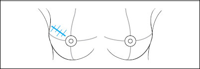
History: Open biopsy of the right breast more than 10 years ago.
Risk profile: No increased risk.
Age: 72 years.
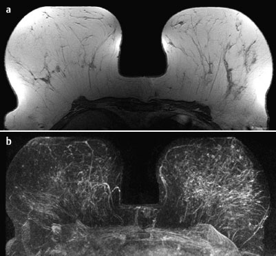
Fig. 76.1 a,b Contrast-enhanced MR mammography. Precontrast image and maximum intensity projection.
Clinical Findings
Normal.
Ultrasound
No unusual findings. (Not shown).
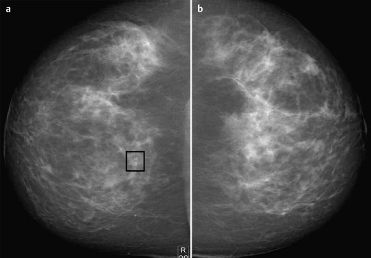
Fig. 76.2a,b Digital mammography, CC view.
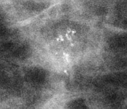
Fig. 76.3 Magnification view (CC) of the central region of the right breast.
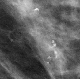
Fig. 76.4 Magnification view (MLO) of the central region of the right breast.

|
Please characterize mammography and MRI findings.
What is your preliminary diagnosis?
What are your next steps? |


