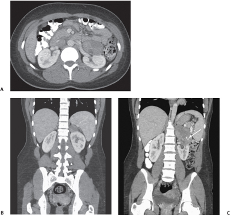
CASE 76 A 20-year-old woman presents with abdominal pain. Fig. 76.1 Left adrenal ganglioneuroma in a 20-year-old woman. (A,B) Axial and coronal enhanced CT scans demonstrate a well-defined oval mass in the left adrenal gland. (C) The mass appears heterogeneous and contains small central punctate calcifications (arrow). A large mass is seen arising from the left adrenal gland with heterogeneous enhancement and calcification (Fig. 76.1). Adrenal ganglioneuroma
Clinical Presentation
Radiologic Findings
Diagnosis
Differential Diagnosis
Discussion
Background
Related posts:


Stay updated, free articles. Join our Telegram channel

Full access? Get Clinical Tree


