VII GI Tract, Peritoneal Cavity, and Retroperitoneum
CASE 77
Clinical Presentation
Dysphagia.
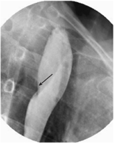
Fig. 77.1 Barium esophagram shows smooth, linear indentation producing a mild filling defect (arrow) over the posterior wall of the esophagus, causing partial obstruction with mild dilatation of the proximal esophagus.
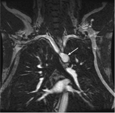
Fig. 77.2 Magnetic resonance angiography image shows the origin (arrow) and course of the aberrant right subclavian artery toward the contralateral side.
Radiologic Findings
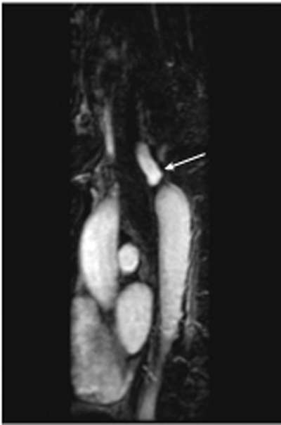
Barium esophagram shows a smooth, linear indentation with regular margins over the posterior wall of the middle third of the esophagus with mild proximal dilatation of the cervical esophagus (Fig. 77.1). Magnetic resonance angiography (MRA) images (Figs. 77.2, 77.3) show an aberrant right subclavian artery (ARSA) arising from the left aortic arch, causing an impression in the posterior wall of the esophagus.
Diagnosis
Aberrant right subclavian artery
Differential Diagnosis
- Double aortic arch
- Esophageal spasm
- Extrinsic compression of the esophagus from nonesophageal tumors (retrosternal extension of the thyroid) or lymphadenopathy
Related posts:


Stay updated, free articles. Join our Telegram channel

Full access? Get Clinical Tree


