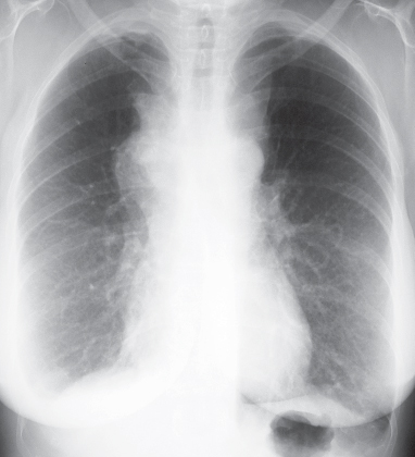
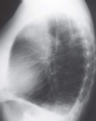
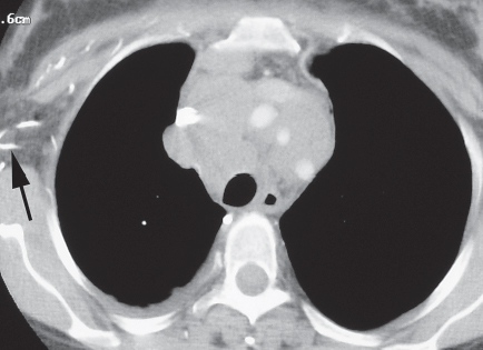
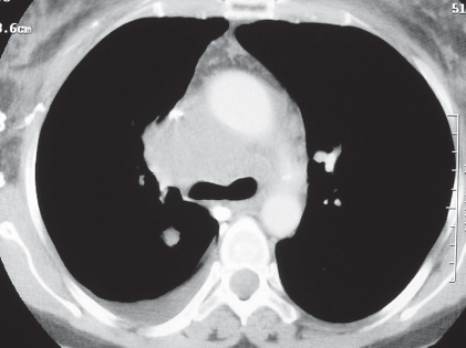
CASE 77 60-year-old woman with facial swelling and weight loss PA (Fig. 77.1) and lateral (Fig. 77.2) chest radiographs demonstrate a mediastinal mass of lobular contours that extends to both sides of midline and is predominantly located in the anterior mediastinum (Fig. 77.2). Note the small right pleural effusion. Contrast-enhanced chest CT (mediastinal window) (Figs. 77.3, 77.4) demonstrates extensive mediastinal lymphadenopathy encasing the great vessels and the central tracheobronchial tree as well as a small right pleural effusion. Note the almost complete obliteration of the lumen of the superior vena cava (Fig. 77.4), enhancing chest wall collateral vessels (arrow) (Fig. 77.3), and intense enhancement of the azygos vein (Fig. 77.4), consistent with superior vena cava obstruction. Lung Cancer; Small Cell Carcinoma • Lung Cancer, Other Cell Type • Lymphoma • Mediastinal Metastases Fig. 77.1 Fig. 77.2 Fig. 77.3 Fig. 77.4
 Clinical Presentation
 Radiologic Findings
 Diagnosis
 Differential Diagnosis
Related posts:
![]()
Stay updated, free articles. Join our Telegram channel

Full access? Get Clinical Tree


Radiology Key
Fastest Radiology Insight Engine


