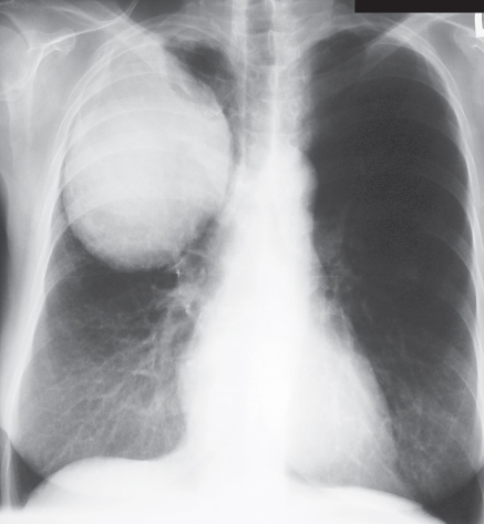
CASE 78 70-year-old woman with right chest wall pain PA (Fig. 78.1) and lateral (Fig. 78.2) chest radiographs demonstrate a large ovoid right upper lobe mass of lobular contours with adjacent right apical pleural thickening and suggestion of destruction of the anterolateral portions of the second and third right ribs. Lung Cancer: Large Cell Carcinoma • Lung Cancer; Other Cell Type • Lymphoma • Lung Abscess Fig. 78.1
 Clinical Presentation
 Radiologic Findings
 Diagnosis
 Differential Diagnosis
Related posts:


Stay updated, free articles. Join our Telegram channel

Full access? Get Clinical Tree


