BI-RADS Categorization | ||
Clinical Findings | right 1 | left 2 |
Ultrasound | right 1 | left 4 |
Mammography | right 1 | left 4 |
BI-RADS Total | right 1 | left 3 |
 Differential Diagnosis
Phyllodes tumor, hamartoma, fibroadenoma, papilloma.
Procedure
Although the lesion was categorized BI-RADS 4, an open biopsy of the lesion was performed without preoperative core biopsy at the request of the patient as well as of her son.
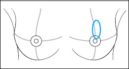
This is the imaging study of a symptomatic woman with a palpable mass discovered a couple of weeks earlier in the center of the left breast. Clinically this lump seemed benign.
Ultrasound
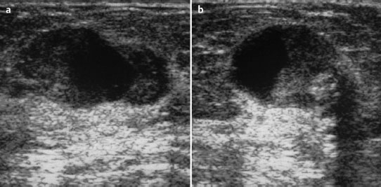
Consistent with the clinical findings, sonography showed a well-defined, mainly solid but partially cystic oval mass in the left breast. Due to the possibility of a malign phyllodes tumor, this was categorized US BI-RADS left 4.
Mammography
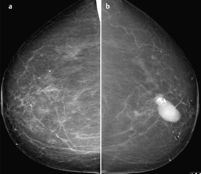
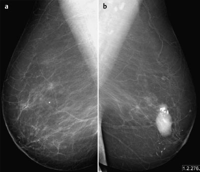
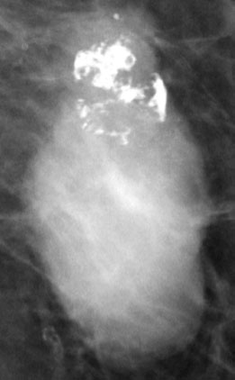
Bilaterally predominantly lipomatous glandular tissue, ACR type 1. Consistent with the palpable finding, there was a well-defined, oval, hyperdense lesion measuring 2 cm with internal popcorn like macrocalcifications in the center of the left breast (Fig. 79.4). Arteriosclerosis. Possibility of a malignant phyllodes tumor adjoining a fibroadenoma with regressive changes; hence categorization as BI-RADS right 1/left 4. PGMICC view M (superimposition of tissue in the right breast laterally, image not repeated); MLO view G (inframammary fold).
Fig. 79.4 Magnification view of the left breast.
Histology
Partially hemorrhagic, partially calcified tumor 17 mm in diameter.
Tumor like sclerosing adenosis.
Stay updated, free articles. Join our Telegram channel

Full access? Get Clinical Tree


