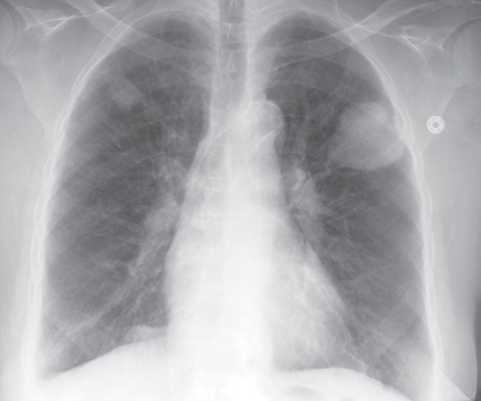
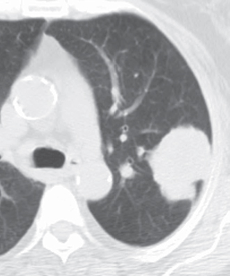
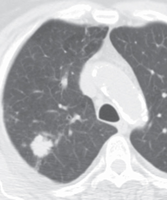
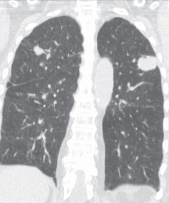
CASE 79 76-year-old woman with chest discomfort and weight loss PA chest radiograph (Fig. 79.1) demonstrates a dominant polylobular mass in the left upper lobe and a polylobular nodule in the right upper lobe. Unenhanced chest CT (lung window) (Figs. 79.2, 79.3) shows the dominant polylobular mass in the left upper lobe (Fig. 79.2) and the right upper lobe polylobular nodule, which exhibits slightly spiculated borders and a pleural tag (Fig. 79.3). Coronal reformatted unenhanced chest CT (lung window) (Fig. 79.4) illustrates the bilateral upper lobe lung cancers. Multicentric Synchronous Lung Cancers • Multifocal Infection • Pulmonary Vasculitis • Pulmonary Metastases; Atypical Manifestation because of Upper Lobe Involvement Fig. 79.1 Fig. 79.2 Fig. 79.3 Fig. 79.4 Multicentric primary lung cancers can be classified as synchronous or metachronous types. Synchronous lung cancers
 Clinical Presentation
 Radiologic Findings
 Diagnosis
 Differential Diagnosis
 Discussion
Background
Related posts:
![]()
Stay updated, free articles. Join our Telegram channel

Full access? Get Clinical Tree


Radiology Key
Fastest Radiology Insight Engine


