
BI-RADS Categorization | ||
Clinical Findings | right 1 | left 1 |
Ultrasound | right 1 | left 1 |
Mammography | right 1 | left 1 |
BI-RADS Total | right 1 | left 1 |
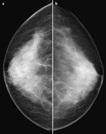
This case presents images from screening mammography.
Ultrasound (not shown)
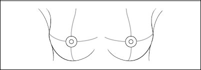
Normal echogenicity of the parenchyma bilaterally. No unusual findings in the right breast behind the nipple or elsewhere. US BI-RADS right 1/left 1.
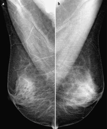
Mammography
Symmetric, inhomogeneously dense parenchyma, ACR type 3. No circumscribed masses or densities. In the area behind the nipple of the right breast there was a lobulated, centrally transparent macrocalcification visible in MLO view. CC view here showed pronounced tramline-like arterial calcifications. No suspicious microcalcifications. BI-RADS right 1/left 1. PGMI: CC view P; MLO view P.
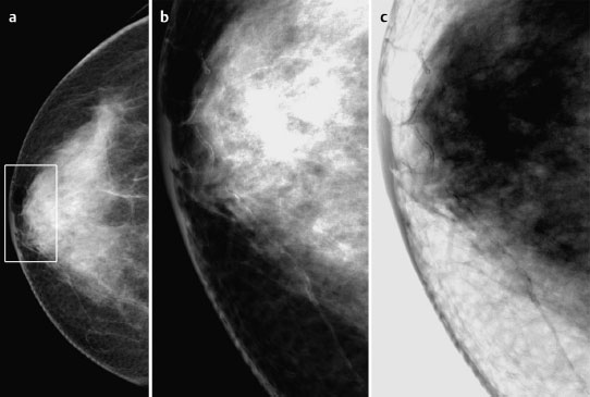
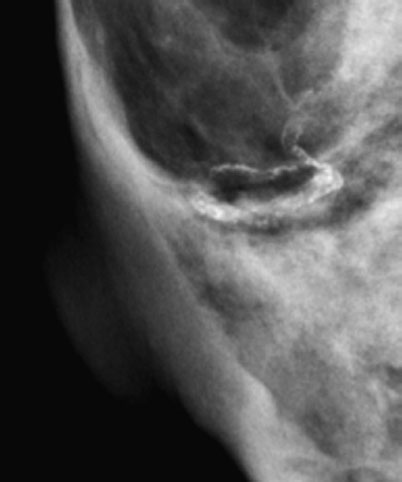
Fig. 80.4a–c Mammography of the right breast in CC view. Enlarged and inverted images.
Diagnosis (no histological confirmation)
Intramammary arteriosclerosis.


Stay updated, free articles. Join our Telegram channel

Full access? Get Clinical Tree


