
Clinical Findings | right 1 | left 1 |
Ultrasound | right 1 | left 1 |
Mammography | right 1 | left 4 |
BI-RADS Total | right 1 | left 4 |
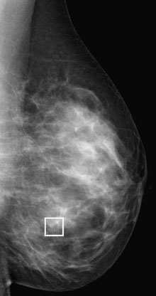
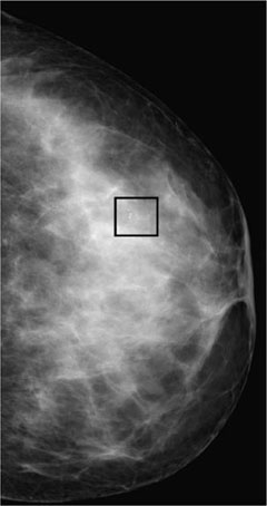
This is a typical screening mammography.
Mammography
ACR type 4 glandular tissue density. In the lower outer quadrant of the left breast at the 4-o’clock position, a cluster of polymorphous microcalcifications could be seen. BI-RADS 4. PGMI is not defined for unilateral mammograms.
Procedure
Histopathological analysis of the microcalcifications with stereotactic vacuum biopsy.
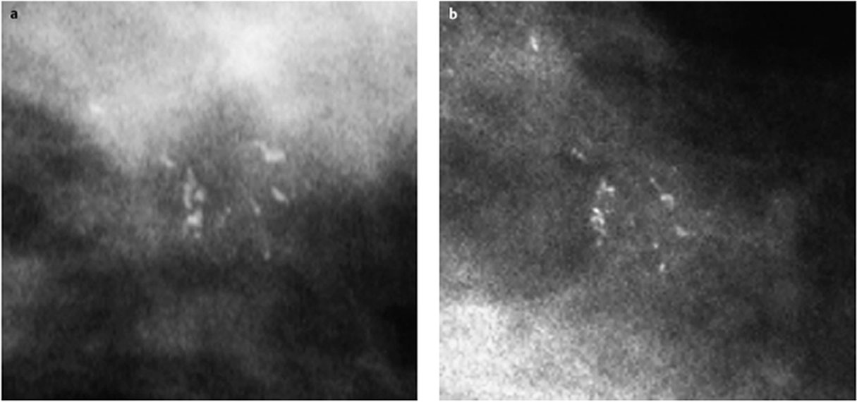
Specimen radiography
Characteristic documentation of calcifications in multiple specimens (Fig. 81.4).
Histopathology of the specimen
Ductal carcinoma in situ (DCIS) (Fig. 81.5).
Procedure
Open biopsy after preoperative localization.
Radiography of the macroscopic specimen
Two residual microcalcifications were depicted near the end of the hook-wire (Fig. 81.6).
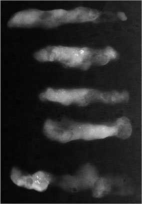
Fig. 81.4 Specimen radiography.
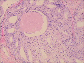
Fig. 81.5 Histology of the specimens.
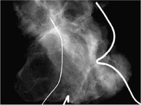
Fig. 81.6 Radiography of the macroscopic specimen.
Histology
DCIS pTis, pNO, MO, RO.

Stay updated, free articles. Join our Telegram channel

Full access? Get Clinical Tree


