Case 81
 Clinical Presentation
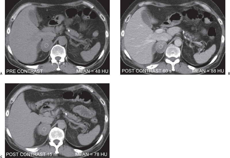
A 68-year-old woman with malignant melanoma. Adrenal washout computed tomography was performed to evaluate an adrenal nodule.
 Imaging Findings
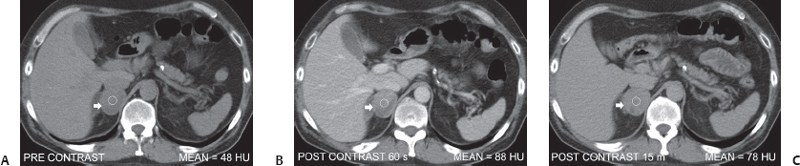
(A) Noncontrast computed tomography (CT) image at the level of the adrenal glands shows a nodule (arrow) in the right adrenal gland. The measured attenuation value is 48 Hounsfield units (HU). (B) CT image obtained 1 minute after the injection of contrast shows heterogeneous enhancement of the adrenal nodule (arrow). The measured attenuation value is 88 HU. (C) CT image obtained 15 minutes after the injection of contrast shows some decrease in enhancement of the nodule (arrow). The measured attenuation value is 78 HU. The estimated absolute washout index is < 60%.
 Differential Diagnosis
• Adrenal metastasis: An attenuation value < 10 HU and a low absolute washout index suggest that the lesion is not an adenoma. Heterogeneous enhancement suggests malignancy. The small size of the nodule, statistically higher frequency of metastases than of primary adrenal carcinomas, and presence of underlying malignant disease favor metastasis over primary adrenal malignancy.
Stay updated, free articles. Join our Telegram channel

Full access? Get Clinical Tree


