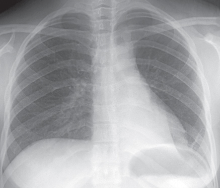
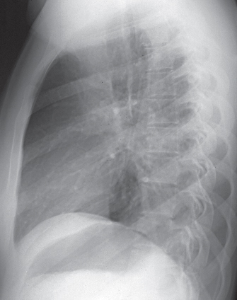
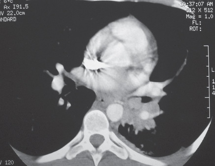
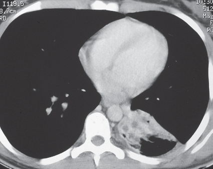
CASE 81 18-year-old woman with cough and hemoptysis PA (Fig. 81.1) and lateral (Fig. 81.2) chest radiographs demonstrate left lower lobe atelectasis. Contrast-enhanced chest CT (mediastinal window) (Figs. 81.3, 81.4) reveals a small, lobular, enhancing, partially endobronchial lesion obstructing the left lower lobe bronchus, with resultant volume loss, consolidation, and bronchiectasis (Fig. 81.4). Bronchial Carcinoid • Mucoepidermoid Carcinoma • Adenoid Cystic Carcinoma • Central Lung Cancer (rare in adolescents and young adults) Fig. 81.1 Fig. 81.2 Fig. 81.3 Fig. 81.4 Carcinoid is a rare malignant primary pulmonary neoplasm of neuroendocrine origin and accounts for approximately 2% of all lung neoplasms. Eighty to 90% of bronchial and pulmonary carcinoids are typical (<2 mitoses per 10 high-power fields and absent necrosis) and 10–20% are atypical (2–10 mitoses per 10 high-power fields and necrosis) carcinoids.
 Clinical Presentation
 Radiologic Findings
 Diagnosis
 Differential Diagnosis
 Discussion
Background
Etiology
Related posts:
![]()
Stay updated, free articles. Join our Telegram channel

Full access? Get Clinical Tree


Radiology Key
Fastest Radiology Insight Engine


