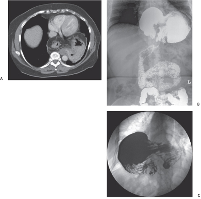
CASE 81 A middle-aged man presents for evaluation of long-standing dysphagia with episodic periods of more intense discomfort. Fig. 81.1 (A) Contrast-enhanced axial CT image demonstrates a large hiatal hernia. The distal esophagus is midline, and the gastroesophageal (GE) junction as well as a portion of the stomach is located within the intrathoracic hernia. (B) Overhead radiograph and (C) spot fluoroscopic image from an upper gastrointestinal series show a large hiatal hernia. The radiograph illustrates that the “concave” greater curvature of the gastric body is located cephalad to the “convex” lesser curvature, the GE junction and pylorus. The GE junction and pylorus maintain their normal relationship. Contrast-enhanced axial computed tomography (CT) image demonstrates a large hiatal hernia (Fig. 81.1A). The distal esophagus is midline, and the gastroesophageal (GE) junction as well as a portion of the stomach is located within the intrathoracic hernia. An overhead radiograph and spot fluoroscopic image (Fig. 81.1B,C) from an upper gastrointestinal (GI) series show a large hiatal hernia. The radiograph illustrates that the “concave” greater curvature of the gastric body is located cephalad to the “convex” lesser curvature, the GE junction and pylorus. The GE junction and pylorus maintain their normal relationship, which excludes a mesenteroaxial gastric volvulus. There is no gastric outlet obstruction. Organoaxial gastric volvulus in a large hiatal hernia Gastric volvulus represents abnormal torsion of the stomach, usually > 180 degrees. If the greater curvature rotates superior to the lesser curvature along the long axis of the stomach, this is termed an organoaxial volvulus and accounts for ~60% of gastric volvulus cases. If the antrum and pylorus rotate anterosuperior to the fundus and GE junction, this is termed a mesenteroaxial volvulus,
Clinical Presentation

Radiologic Findings
Diagnosis
Differential Diagnosis
Discussion
Background
![]()
Stay updated, free articles. Join our Telegram channel

Full access? Get Clinical Tree


