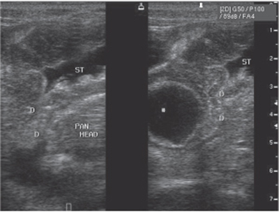
CASE 82 A 42-year-old man presents with postprandial epigastric discomfort. Fig. 82.1 Ultrasound image shows a well-defined cystic structure with a gut signature in continuity with the lateral duodenal wall and the lumen of the vertical part of the duodenum. The lumen and the wall of the lesion are contiguous with the lateral wall of the second part of the duodenum. Upper abdominal ultrasound shows a well-demarcated hypoechoic structure (Fig. 82.1) in close proximity to the second portion of the duodenum. The structure has a gut signature on ultrasound with alternating hyper- and hypoechoic layers in the wall. Duodenal diverticulum arising from the lateral wall of the duodenum The duodenum is the second most common site for diverticula formation after the large bowel. The incidence of duodenal diverticula is 5 to 15% on barium examinations, 20 to 25% at autopsy. The peak incidence of duodenal diverticula is found in the 5th to 6th decades of life. They typically arise from the medial wall of the second and third parts of the duodenum within 2 cm of the ampulla of Vater. Intraluminal duodenal diverticula are rare. Diverticula arising from the lateral wall tend to be larger in size and are less common. Diverticula can also be classified as primary or secondary (due to postbulbar ulcer).
Clinical Presentation
Radiologic Findings
Diagnosis
Differential Diagnosis
Discussion
Background
Clinical Findings
Related posts:


Stay updated, free articles. Join our Telegram channel

Full access? Get Clinical Tree


